


 Foundational characteristics of cancer include proliferation, angiogenesis, migration, evasion of apoptosis, and cellular immortality. Find key markers for these cellular processes and antibodies to detect them.
 The SUMOplot™ Analysis Program predicts and scores sumoylation sites in your protein. SUMOylation is a post-translational modification involved in various cellular processes, such as nuclear-cytosolic transport, transcriptional regulation, apoptosis, protein stability, response to stress, and progression through the cell cycle.
 The Autophagy Receptor Motif Plotter predicts and scores autophagy receptor binding sites in your protein. Identifying proteins connected to this pathway is critical to understanding the role of autophagy in physiological as well as pathological processes such as development, differentiation, neurodegenerative diseases, stress, infection, and cancer.
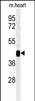
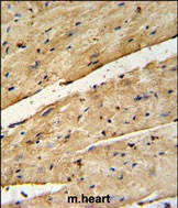
B3GNT7 Antibody (Center)
Affinity Purified Rabbit Polyclonal Antibody (Pab)
- SPECIFICATION
- CITATIONS
- PROTOCOLS
- BACKGROUND

Application
| IHC-P, WB, E |
|---|---|
| Primary Accession | Q8NFL0 |
| Reactivity | Human, Mouse |
| Host | Rabbit |
| Clonality | Polyclonal |
| Isotype | Rabbit IgG |
| Calculated MW | 45987 Da |
| Antigen Region | 256-282 aa |
| Gene ID | 93010 |
|---|---|
| Other Names | UDP-GlcNAc:betaGal beta-1, 3-N-acetylglucosaminyltransferase 7, BGnT-7, Beta-1, 3-Gn-T7, Beta-1, 3-N-acetylglucosaminyltransferase 7, Beta3Gn-T7, 241-, B3GNT7 |
| Target/Specificity | This B3GNT7 antibody is generated from rabbits immunized with a KLH conjugated synthetic peptide between 256-282 amino acids from the Central region of human B3GNT7. |
| Dilution | IHC-P~~1:50~100 WB~~1:1000 E~~Use at an assay dependent concentration. |
| Format | Purified polyclonal antibody supplied in PBS with 0.09% (W/V) sodium azide. This antibody is purified through a protein A column, followed by peptide affinity purification. |
| Storage | Maintain refrigerated at 2-8°C for up to 2 weeks. For long term storage store at -20°C in small aliquots to prevent freeze-thaw cycles. |
| Precautions | B3GNT7 Antibody (Center) is for research use only and not for use in diagnostic or therapeutic procedures. |
| Name | B3GNT7 {ECO:0000303|PubMed:17690104, ECO:0000312|HGNC:HGNC:18811} |
|---|---|
| Function | N-acetyl glucosamine (GlcNAc) transferase that catalyzes the transfer of GlcNAc via a beta1->3 linkage from UDP-GlcNAc to the non- reducing terminal galactose (Gal) in the linearly growing chain of N- and O-linked keratan sulfate proteoglycans. Cooperates with B4GALT4 galactosyltransferase and CHST6 and CHST1 sulfotransferases to construct and elongate mono- and disulfated disaccharide units [->3Galbeta1->4(6-sulfoGlcNAcbeta)1->] and [->3(6-sulfoGalbeta)1->4(6- sulfoGlcNAcbeta)1->] within keratan sulfate polymer (PubMed:14706853, PubMed:17690104). Involved in biosynthesis of N-linked keratan sulfate proteoglycans in cornea, with an impact on proteoglycan fibril organization and corneal transparency (By similarity) (PubMed:17690104). May play a role in the maintenance of tissue architecture by suppressing cellular motility and invasion (By similarity). |
| Cellular Location | Golgi apparatus membrane; Single- pass type II membrane protein |
| Tissue Location | Expressed in corneal epithelial cells. |

Thousands of laboratories across the world have published research that depended on the performance of antibodies from Abcepta to advance their research. Check out links to articles that cite our products in major peer-reviewed journals, organized by research category.
info@abcepta.com, and receive a free "I Love Antibodies" mug.

Provided below are standard protocols that you may find useful for product applications.
Background
B3GNT7 is a glycosyltransferase which may play a role in elongation of the keratan sulfate carbohydrate backbone.
References
Kitayama, K., et al. J. Biol. Chem. 282(41):30085-30096(2007)
Seko, A., et al. FEBS Lett. 556 (1-3), 216-220 (2004)
Kataoka, K., et al. Biochem. Biophys. Res. Commun. 294(4):843-848(2002)
If you have used an Abcepta product and would like to share how it has performed, please click on the "Submit Review" button and provide the requested information. Our staff will examine and post your review and contact you if needed.
If you have any additional inquiries please email technical services at tech@abcepta.com.
Ordering Information
Other Products
Shipping Information
