


 Foundational characteristics of cancer include proliferation, angiogenesis, migration, evasion of apoptosis, and cellular immortality. Find key markers for these cellular processes and antibodies to detect them.
 The SUMOplot™ Analysis Program predicts and scores sumoylation sites in your protein. SUMOylation is a post-translational modification involved in various cellular processes, such as nuclear-cytosolic transport, transcriptional regulation, apoptosis, protein stability, response to stress, and progression through the cell cycle.
 The Autophagy Receptor Motif Plotter predicts and scores autophagy receptor binding sites in your protein. Identifying proteins connected to this pathway is critical to understanding the role of autophagy in physiological as well as pathological processes such as development, differentiation, neurodegenerative diseases, stress, infection, and cancer.
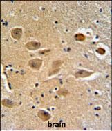
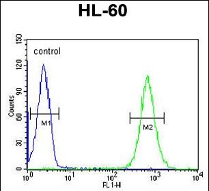
MILK2 Antibody (C-term)
Affinity Purified Rabbit Polyclonal Antibody (Pab)

- SPECIFICATION
- CITATIONS
- PROTOCOLS
- BACKGROUND

Application
| FC, IHC-P, WB, E |
|---|---|
| Primary Accession | Q8IY33 |
| Reactivity | Human |
| Host | Rabbit |
| Clonality | Polyclonal |
| Isotype | Rabbit IgG |
| Calculated MW | 97502 Da |
| Antigen Region | 841-870 aa |
| Gene ID | 79778 |
|---|---|
| Other Names | MICAL-like protein 2, Junctional Rab13-binding protein, Molecule interacting with CasL-like 2, MICAL-L2, MICALL2, JRAB |
| Target/Specificity | This MILK2 antibody is generated from rabbits immunized with a KLH conjugated synthetic peptide between 841-870 amino acids from the C-terminal region of human MILK2. |
| Dilution | FC~~1:10~50 IHC-P~~1:50~100 WB~~1:1000 E~~Use at an assay dependent concentration. |
| Format | Purified polyclonal antibody supplied in PBS with 0.09% (W/V) sodium azide. This antibody is purified through a protein A column, followed by peptide affinity purification. |
| Storage | Maintain refrigerated at 2-8°C for up to 2 weeks. For long term storage store at -20°C in small aliquots to prevent freeze-thaw cycles. |
| Precautions | MILK2 Antibody (C-term) is for research use only and not for use in diagnostic or therapeutic procedures. |
| Name | MICALL2 (HGNC:29672) |
|---|---|
| Synonyms | JRAB |
| Function | Effector of small Rab GTPases which is involved in junctional complexes assembly through the regulation of cell adhesion molecules transport to the plasma membrane and actin cytoskeleton reorganization. Regulates the endocytic recycling of occludins, claudins and E-cadherin to the plasma membrane and may thereby regulate the establishment of tight junctions and adherens junctions. In parallel, may regulate actin cytoskeleton reorganization directly through interaction with F-actin or indirectly through actinins and filamins. Most probably involved in the processes of epithelial cell differentiation, cell spreading and neurite outgrowth (By similarity). Undergoes liquid-liquid phase separation to form tubular recycling endosomes. Plays 2 sequential roles in the biogenesis of tubular recycling endosomes: first organizes phase separation and then the closed form formed by interaction with RAB8A promotes endosomal tubulation (By similarity). |
| Cellular Location | Cell membrane {ECO:0000250|UniProtKB:Q3TN34}; Peripheral membrane protein {ECO:0000250|UniProtKB:Q3TN34}. Cell junction, tight junction {ECO:0000250|UniProtKB:Q3TN34}. Recycling endosome {ECO:0000250|UniProtKB:Q3TN34}. Cell projection {ECO:0000250|UniProtKB:Q3TN34}. Cytoplasm, cytoskeleton {ECO:0000250|UniProtKB:Q3TN34} |

Thousands of laboratories across the world have published research that depended on the performance of antibodies from Abcepta to advance their research. Check out links to articles that cite our products in major peer-reviewed journals, organized by research category.
info@abcepta.com, and receive a free "I Love Antibodies" mug.

Provided below are standard protocols that you may find useful for product applications.
Background
MILK2 may be a cytoskeletal regulator.
References
Kanda, I., et al. Oncogene 27(12):1687-1695(2008)
Terai, T., et al. Mol. Biol. Cell 17(5):2465-2475(2006)
Clark, H.F., et al. Genome Res. 13(10):2265-2270(2003)
Terman, J.R., et al. Cell 109(7):887-900(2002)
If you have used an Abcepta product and would like to share how it has performed, please click on the "Submit Review" button and provide the requested information. Our staff will examine and post your review and contact you if needed.
If you have any additional inquiries please email technical services at tech@abcepta.com.
Ordering Information
Other Products
Shipping Information
