


 Foundational characteristics of cancer include proliferation, angiogenesis, migration, evasion of apoptosis, and cellular immortality. Find key markers for these cellular processes and antibodies to detect them.
 The SUMOplot™ Analysis Program predicts and scores sumoylation sites in your protein. SUMOylation is a post-translational modification involved in various cellular processes, such as nuclear-cytosolic transport, transcriptional regulation, apoptosis, protein stability, response to stress, and progression through the cell cycle.
 The Autophagy Receptor Motif Plotter predicts and scores autophagy receptor binding sites in your protein. Identifying proteins connected to this pathway is critical to understanding the role of autophagy in physiological as well as pathological processes such as development, differentiation, neurodegenerative diseases, stress, infection, and cancer.
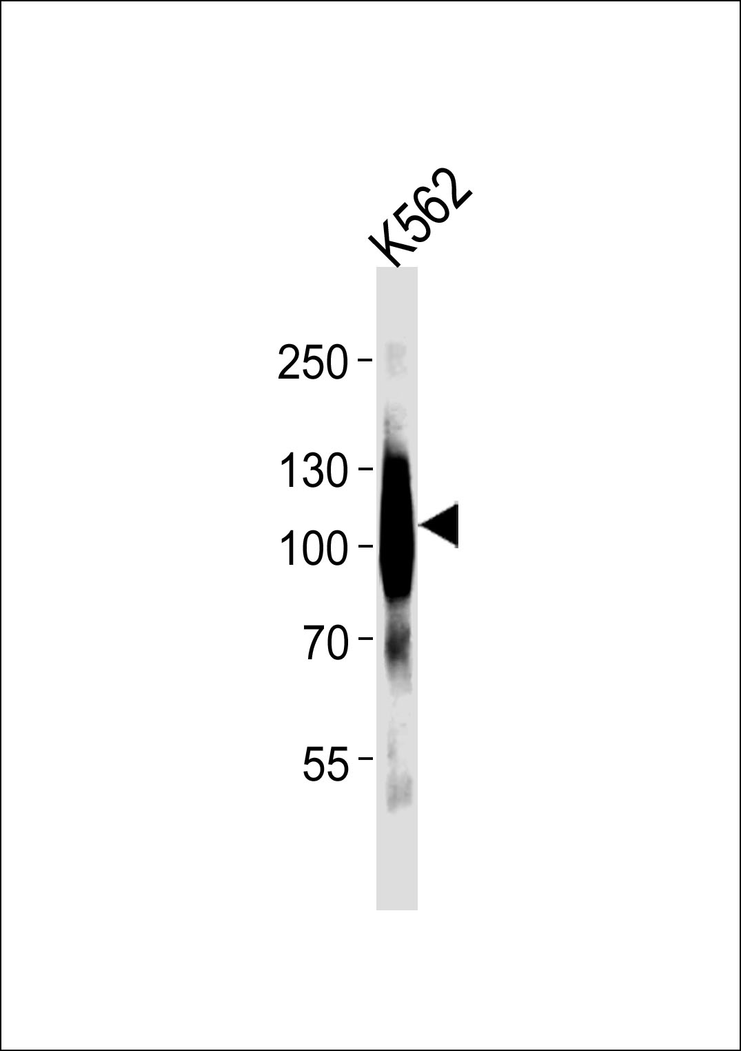
mGluR6 Antibody
Purified Rabbit Polyclonal Antibody (Pab)
- SPECIFICATION
- CITATIONS
- PROTOCOLS
- BACKGROUND

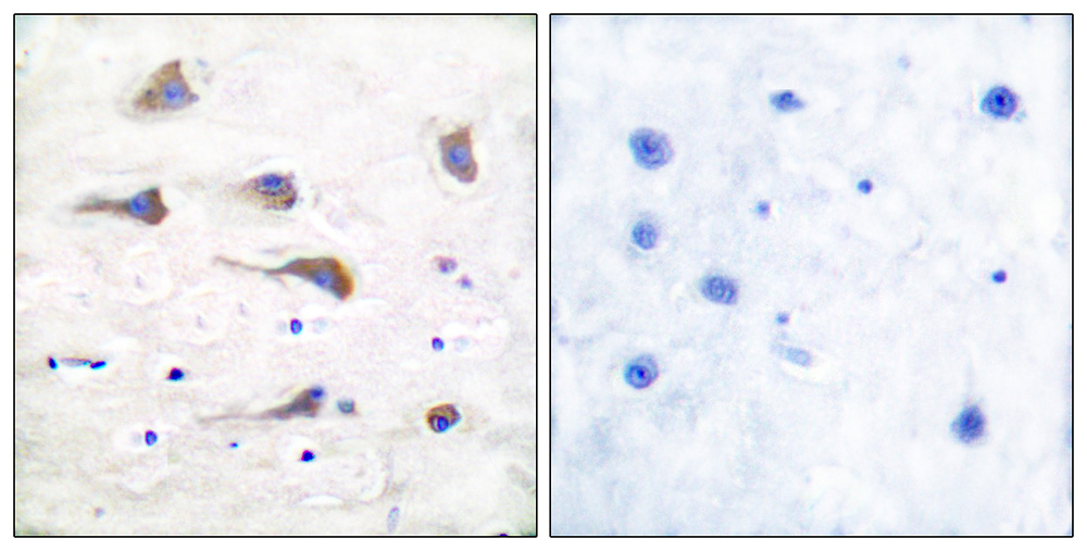
Application
| WB, IHC |
|---|---|
| Primary Accession | O15303 |
| Reactivity | Human |
| Host | Rabbit |
| Clonality | Polyclonal |
| Calculated MW | 95 KDa |
| Antigen Region | 850-877 aa |
| Gene ID | 2916 |
|---|---|
| Other Names | Metabotropic glutamate receptor 6, mGluR6, GRM6, GPRC1F, MGLUR6 |
| Dilution | WB~~ 1:1000 IHC~~1:50-1:100 |
| Format | Rabbit IgG in phosphate buffered saline (without Mg2+ and Ca2+), pH 7.4, 150mM NaCl, 0.09% (W/V) sodium azide and 50% glycerol. |
| Storage Conditions | -20℃ |
| Name | GRM6 |
|---|---|
| Synonyms | GPRC1F, MGLUR6 |
| Function | G-protein coupled receptor for glutamate. Ligand binding causes a conformation change that triggers signaling via guanine nucleotide-binding proteins (G proteins) and modulates the activity of down-stream effectors, such as adenylate cyclase. Signaling inhibits adenylate cyclase activity (By similarity). Signaling stimulates TRPM1 channel activity and Ca(2+) uptake. Required for normal vision. |
| Cellular Location | Cell membrane; Multi-pass membrane protein. Endoplasmic reticulum membrane; Multi-pass membrane protein. Golgi apparatus membrane; Multi-pass membrane protein. Cell projection, dendrite Note=Subject to trafficking from the endoplasmic reticulum to the Golgi apparatus and then to the cell membrane |
| Tissue Location | Detected in melanocytes. |

Thousands of laboratories across the world have published research that depended on the performance of antibodies from Abcepta to advance their research. Check out links to articles that cite our products in major peer-reviewed journals, organized by research category.
info@abcepta.com, and receive a free "I Love Antibodies" mug.

Provided below are standard protocols that you may find useful for product applications.
Background
G-protein coupled receptor for glutamate. Ligand binding causes a conformation change that triggers signaling via guanine nucleotide-binding proteins (G proteins) and modulates the activity of down-stream effectors, such as adenylate cyclase. Signaling inhibits adenylate cyclase activity (By similarity). Signaling stimulates TRPM1 channel activity and Ca(2+) uptake. Required for normal vision.
References
Hashimoto T.,et al.Eur. J. Neurosci. 9:1226-1235(1997).
Schmutz J.,et al.Nature 431:268-274(2004).
Devi S.,et al.Pigment Cell Melanoma Res. 26:348-356(2013).
Dryja T.P.,et al.Proc. Natl. Acad. Sci. U.S.A. 102:4884-4889(2005).
Sjoeblom T.,et al.Science 314:268-274(2006).
If you have used an Abcepta product and would like to share how it has performed, please click on the "Submit Review" button and provide the requested information. Our staff will examine and post your review and contact you if needed.
If you have any additional inquiries please email technical services at tech@abcepta.com.
Ordering Information
Other Products
Shipping Information
