


 Foundational characteristics of cancer include proliferation, angiogenesis, migration, evasion of apoptosis, and cellular immortality. Find key markers for these cellular processes and antibodies to detect them.
 The SUMOplot™ Analysis Program predicts and scores sumoylation sites in your protein. SUMOylation is a post-translational modification involved in various cellular processes, such as nuclear-cytosolic transport, transcriptional regulation, apoptosis, protein stability, response to stress, and progression through the cell cycle.
 The Autophagy Receptor Motif Plotter predicts and scores autophagy receptor binding sites in your protein. Identifying proteins connected to this pathway is critical to understanding the role of autophagy in physiological as well as pathological processes such as development, differentiation, neurodegenerative diseases, stress, infection, and cancer.
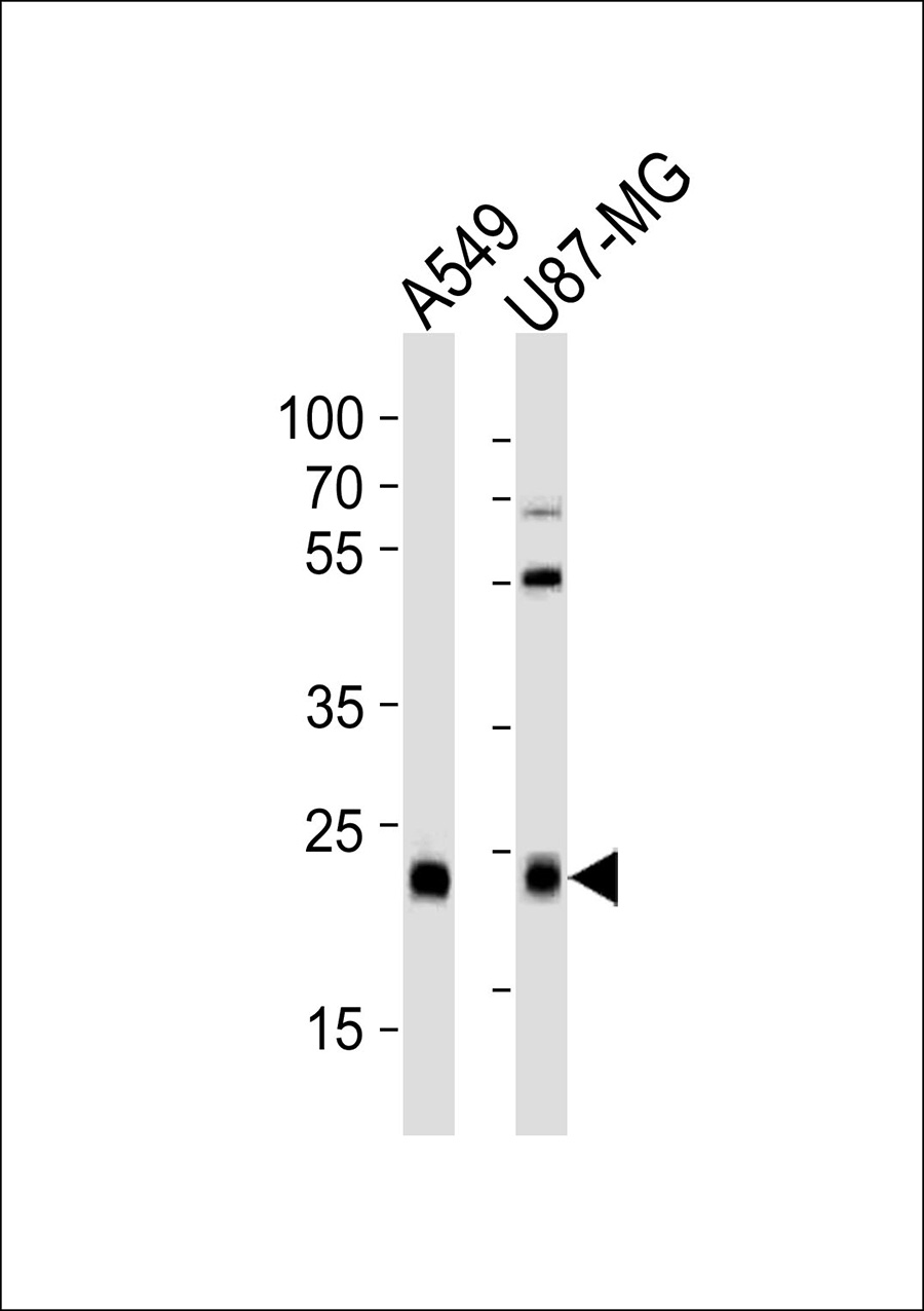
CATL1 Antibody (heavy chain, Cleaved-Thr288)
Purified Rabbit Polyclonal Antibody (Pab)
- SPECIFICATION
- CITATIONS
- PROTOCOLS
- BACKGROUND

Application
| WB |
|---|---|
| Primary Accession | P07711 |
| Reactivity | Human |
| Host | Rabbit |
| Clonality | Polyclonal |
| Calculated MW | 38 KDa |
| Antigen Region | 275-295 aa |
| Gene ID | 1514 |
|---|---|
| Other Names | Cathepsin L1, Cathepsin L, Major excreted protein, MEP, Cathepsin L1 heavy chain, Cathepsin L1 light chain, CTSL, CTSL1 |
| Dilution | WB~~1:1000 |
| Format | Rabbit IgG in phosphate buffered saline (without Mg2+ and Ca2+), pH 7.4, 150mM NaCl, 0.09% (W/V) sodium azide and 50% glycerol. |
| Storage Conditions | -20℃ |
| Name | CTSL (HGNC:2537) |
|---|---|
| Synonyms | CTSL1 |
| Function | Thiol protease important for the overall degradation of proteins in lysosomes (Probable). Plays a critical for normal cellular functions such as general protein turnover, antigen processing and bone remodeling. Involved in the solubilization of cross-linked TG/thyroglobulin and in the subsequent release of thyroid hormone thyroxine (T4) by limited proteolysis of TG/thyroglobulin in the thyroid follicle lumen (By similarity). In neuroendocrine chromaffin cells secretory vesicles, catalyzes the prohormone proenkephalin processing to the active enkephalin peptide neurotransmitter (By similarity). In thymus, regulates CD4(+) T cell positive selection by generating the major histocompatibility complex class II (MHCII) bound peptide ligands presented by cortical thymic epithelial cells. Also mediates invariant chain processing in cortical thymic epithelial cells (By similarity). Major elastin-degrading enzyme at neutral pH. Accumulates as a mature and active enzyme in the extracellular space of antigen presenting cells (APCs) to regulate degradation of the extracellular matrix in the course of inflammation (By similarity). Secreted form generates endostatin from COL18A1 (PubMed:10716919). Critical for cardiac morphology and function. Plays an important role in hair follicle morphogenesis and cycling, as well as epidermal differentiation (By similarity). Required for maximal stimulation of steroidogenesis by TIMP1 (By similarity). |
| Cellular Location | Lysosome {ECO:0000250|UniProtKB:P06797}. Apical cell membrane {ECO:0000250|UniProtKB:P06797}; Peripheral membrane protein {ECO:0000250|UniProtKB:P06797}; Extracellular side {ECO:0000250|UniProtKB:P06797}. Cytoplasmic vesicle, secretory vesicle, chromaffin granule {ECO:0000250|UniProtKB:P25975}. Secreted, extracellular space {ECO:0000250|UniProtKB:P06797}. Secreted {ECO:0000250|UniProtKB:P06797}. Note=Localizes to the apical membrane of thyroid epithelial cells. Released at extracellular space by activated dendritic cells and macrophages {ECO:0000250|UniProtKB:P06797} |

Thousands of laboratories across the world have published research that depended on the performance of antibodies from Abcepta to advance their research. Check out links to articles that cite our products in major peer-reviewed journals, organized by research category.
info@abcepta.com, and receive a free "I Love Antibodies" mug.

Provided below are standard protocols that you may find useful for product applications.
Background
Important for the overall degradation of proteins in lysosomes.
References
Gal S.,et al.Biochem. J. 253:303-306(1988).
Joseph L.J.,et al.J. Clin. Invest. 81:1621-1629(1988).
Ebert L.,et al.Submitted (JUN-2004) to the EMBL/GenBank/DDBJ databases.
Bechtel S.,et al.BMC Genomics 8:399-399(2007).
Humphray S.J.,et al.Nature 429:369-374(2004).
If you have used an Abcepta product and would like to share how it has performed, please click on the "Submit Review" button and provide the requested information. Our staff will examine and post your review and contact you if needed.
If you have any additional inquiries please email technical services at tech@abcepta.com.
Ordering Information
Other Products
Shipping Information
