


 Foundational characteristics of cancer include proliferation, angiogenesis, migration, evasion of apoptosis, and cellular immortality. Find key markers for these cellular processes and antibodies to detect them.
 The SUMOplot™ Analysis Program predicts and scores sumoylation sites in your protein. SUMOylation is a post-translational modification involved in various cellular processes, such as nuclear-cytosolic transport, transcriptional regulation, apoptosis, protein stability, response to stress, and progression through the cell cycle.
 The Autophagy Receptor Motif Plotter predicts and scores autophagy receptor binding sites in your protein. Identifying proteins connected to this pathway is critical to understanding the role of autophagy in physiological as well as pathological processes such as development, differentiation, neurodegenerative diseases, stress, infection, and cancer.
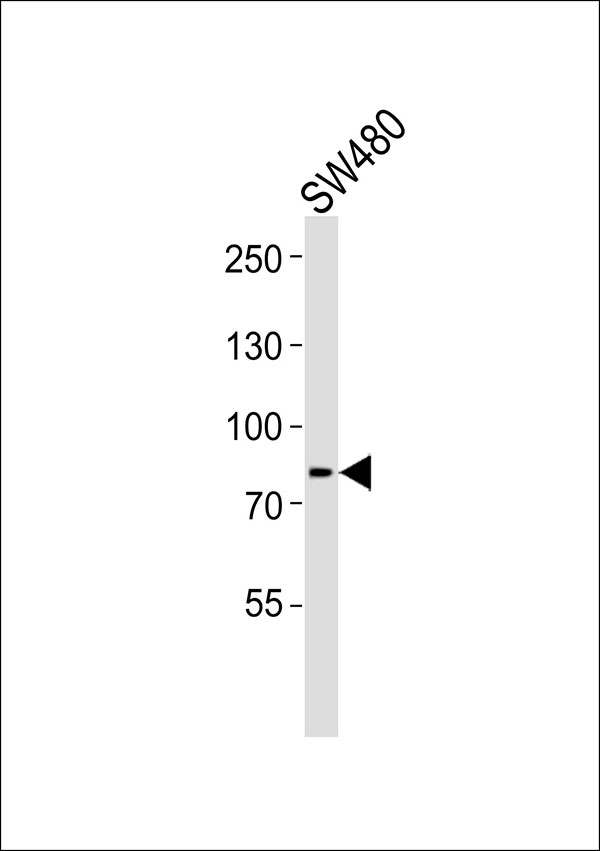
OXR1 Antibody
Purified Rabbit Polyclonal Antibody (Pab)
- SPECIFICATION
- CITATIONS
- PROTOCOLS
- BACKGROUND

Application
| WB |
|---|---|
| Primary Accession | Q8N573 |
| Reactivity | Human, Mouse, Rat |
| Host | Rabbit |
| Clonality | Polyclonal |
| Calculated MW | 98,94,56,28,95,34,24 KDa |
| Antigen Region | 573-599 aa |
| Gene ID | 55074 |
|---|---|
| Other Names | Oxidation resistance protein 1, OXR1 |
| Dilution | WB~~1:1000 |
| Format | Rabbit IgG in phosphate buffered saline (without Mg2+ and Ca2+), pH 7.4, 150mM NaCl, 0.09% (W/V) sodium azide and 50% glycerol. This antibody is purified through a protein A column, followed by peptide affinity purification. |
| Storage Conditions | -20℃ |
| Name | OXR1 |
|---|---|
| Function | May be involved in protection from oxidative damage. |
| Cellular Location | Mitochondrion. |

Thousands of laboratories across the world have published research that depended on the performance of antibodies from Abcepta to advance their research. Check out links to articles that cite our products in major peer-reviewed journals, organized by research category.
info@abcepta.com, and receive a free "I Love Antibodies" mug.

Provided below are standard protocols that you may find useful for product applications.
Background
May be involved in protection from oxidative damage.
References
Yang M.,et al.Submitted (JAN-2010) to the EMBL/GenBank/DDBJ databases.
Ohira M.,et al.Cancer Lett. 197:63-68(2003).
Ota T.,et al.Nat. Genet. 36:40-45(2004).
Nusbaum C.,et al.Nature 439:331-335(2006).
Bechtel S.,et al.BMC Genomics 8:399-399(2007).
If you have used an Abcepta product and would like to share how it has performed, please click on the "Submit Review" button and provide the requested information. Our staff will examine and post your review and contact you if needed.
If you have any additional inquiries please email technical services at tech@abcepta.com.
Ordering Information
Other Products
Shipping Information
