


 Foundational characteristics of cancer include proliferation, angiogenesis, migration, evasion of apoptosis, and cellular immortality. Find key markers for these cellular processes and antibodies to detect them.
 The SUMOplot™ Analysis Program predicts and scores sumoylation sites in your protein. SUMOylation is a post-translational modification involved in various cellular processes, such as nuclear-cytosolic transport, transcriptional regulation, apoptosis, protein stability, response to stress, and progression through the cell cycle.
 The Autophagy Receptor Motif Plotter predicts and scores autophagy receptor binding sites in your protein. Identifying proteins connected to this pathway is critical to understanding the role of autophagy in physiological as well as pathological processes such as development, differentiation, neurodegenerative diseases, stress, infection, and cancer.
Galectin 1 Antibody
Purified Rabbit Polyclonal Antibody (Pab)
- SPECIFICATION
- CITATIONS
- PROTOCOLS
- BACKGROUND

Application
| WB |
|---|---|
| Primary Accession | P09382 |
| Reactivity | Human, Mouse, Rat |
| Host | Rabbit |
| Clonality | Polyclonal |
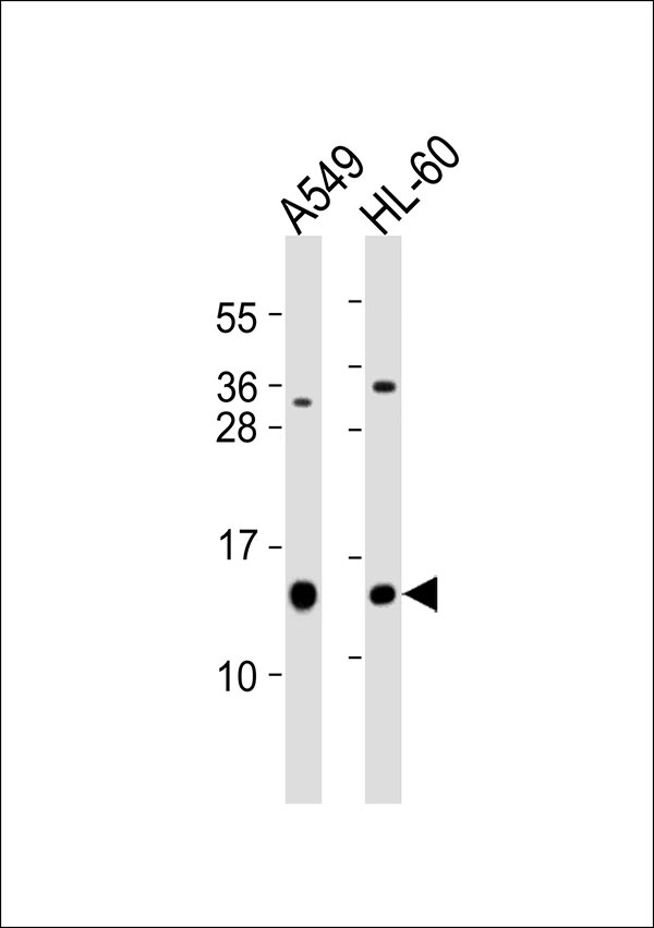
| Calculated MW | 15 KDa |
| Antigen Region | 31 - 90 aa |
| Gene ID | 3956 |
|---|---|
| Other Names | Galectin-1, Gal-1, 14 kDa laminin-binding protein, HLBP14, 14 kDa lectin, Beta-galactoside-binding lectin L-14-I, Galaptin, HBL, HPL, Lactose-binding lectin 1, Lectin galactoside-binding soluble 1, Putative MAPK-activating protein PM12, S-Lac lectin 1, LGALS1 |
| Target/Specificity | KLH-conjugated synthetic peptide encompassing a sequence within the center region of human Galectin 1. The exact sequence is proprietary. |
| Dilution | WB~~ 1:1000 |
| Format | 0.01M PBS, pH 7.2, 0.09% (W/V) Sodium azide, Glycerol 50% |
| Storage | Store at -20 °C.Stable for 12 months from date of receipt |
| Name | LGALS1 (HGNC:6561) |
|---|---|
| Function | Lectin that binds beta-galactoside and a wide array of complex carbohydrates. Plays a role in regulating apoptosis, cell proliferation and cell differentiation. Inhibits CD45 protein phosphatase activity and therefore the dephosphorylation of Lyn kinase. Strong inducer of T-cell apoptosis. Plays a negative role in Th17 cell differentiation via activation of the receptor CD69 (PubMed:24752896). |
| Cellular Location | Secreted, extracellular space, extracellular matrix. Cytoplasm. Secreted Note=Can be secreted; the secretion is dependent on protein unfolding and facilitated by the cargo receptor TMED10; it results in protein translocation from the cytoplasm into the ERGIC (endoplasmic reticulum- Golgi intermediate compartment) followed by vesicle entry and secretion. |
| Tissue Location | Expressed in placenta, maternal decidua and fetal membranes. Within placenta, expressed in trophoblasts, stromal cells, villous endothelium, syncytiotrophoblast apical membrane and villous stroma. Within fetal membranes, expressed in amnion, chorioamniotic mesenchyma and chorion (at protein level). Expressed in cardiac, smooth, and skeletal muscle, neurons, thymus, kidney and hematopoietic cells. |

Thousands of laboratories across the world have published research that depended on the performance of antibodies from Abcepta to advance their research. Check out links to articles that cite our products in major peer-reviewed journals, organized by research category.
info@abcepta.com, and receive a free "I Love Antibodies" mug.

Provided below are standard protocols that you may find useful for product applications.
Background
May regulate apoptosis, cell proliferation and cell differentiation. Binds beta-galactoside and a wide array of complex carbohydrates. Inhibits CD45 protein phosphatase activity and therefore the dephosphorylation of Lyn kinase. Strong inducer of T-cell apoptosis.
References
Hirabayashi J.,et al.Biochim. Biophys. Acta 1008:85-91(1989).
Couraud P.-O.,et al.J. Biol. Chem. 264:1310-1316(1989).
Abbott W.M.,et al.Biochem. J. 259:291-294(1989).
Than N.G.,et al.Proc. Natl. Acad. Sci. U.S.A. 105:15819-15824(2008).
Gitt M.A.,et al.Biochemistry 30:82-89(1991).
If you have used an Abcepta product and would like to share how it has performed, please click on the "Submit Review" button and provide the requested information. Our staff will examine and post your review and contact you if needed.
If you have any additional inquiries please email technical services at tech@abcepta.com.
Ordering Information
Other Products
Shipping Information
