


 Foundational characteristics of cancer include proliferation, angiogenesis, migration, evasion of apoptosis, and cellular immortality. Find key markers for these cellular processes and antibodies to detect them.
 The SUMOplot™ Analysis Program predicts and scores sumoylation sites in your protein. SUMOylation is a post-translational modification involved in various cellular processes, such as nuclear-cytosolic transport, transcriptional regulation, apoptosis, protein stability, response to stress, and progression through the cell cycle.
 The Autophagy Receptor Motif Plotter predicts and scores autophagy receptor binding sites in your protein. Identifying proteins connected to this pathway is critical to understanding the role of autophagy in physiological as well as pathological processes such as development, differentiation, neurodegenerative diseases, stress, infection, and cancer.
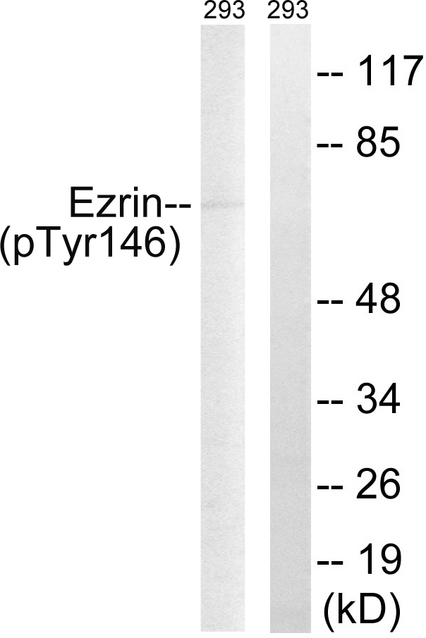
Ezrin (Phospho-Tyr146) Antibody
Purified Rabbit Polyclonal Antibody (Pab)
- SPECIFICATION
- CITATIONS
- PROTOCOLS
- BACKGROUND

Application
| WB |
|---|---|
| Primary Accession | P15311 |
| Reactivity | Human, Mouse, Rat |
| Host | Rabbit |
| Clonality | Polyclonal |
| Calculated MW | 69413 Da |
| Gene ID | 7430 |
|---|---|
| Other Names | Ezrin, Cytovillin, Villin-2, p81, EZR, VIL2 |
| Dilution | WB~~1:1000 |
| Format | Rabbit IgG in phosphate buffered saline (without Mg2+ and Ca2+), pH 7.4, 150mM NaCl, 0.09% (W/V) sodium azide and 50% glycerol. |
| Storage Conditions | -20℃ |
| Name | EZR |
|---|---|
| Synonyms | VIL2 |
| Function | Probably involved in connections of major cytoskeletal structures to the plasma membrane. In epithelial cells, required for the formation of microvilli and membrane ruffles on the apical pole. Along with PLEKHG6, required for normal macropinocytosis. |
| Cellular Location | Apical cell membrane; Peripheral membrane protein; Cytoplasmic side. Cell projection. Cell projection, microvillus membrane; Peripheral membrane protein; Cytoplasmic side. Cell projection, ruffle membrane; Peripheral membrane protein; Cytoplasmic side. Cytoplasm, cell cortex. Cytoplasm, cytoskeleton. Cell projection, microvillus {ECO:0000250|UniProtKB:P26040}. Note=Localization to the apical membrane of parietal cells depends on the interaction with PALS1 Localizes to cell extensions and peripheral processes of astrocytes (By similarity). Microvillar peripheral membrane protein (cytoplasmic side). {ECO:0000250|UniProtKB:P31977} |
| Tissue Location | Expressed in cerebral cortex, basal ganglia, hippocampus, hypophysis, and optic nerve. Weakly expressed in brain stem and diencephalon. Stronger expression was detected in gray matter of frontal lobe compared to white matter (at protein level). Component of the microvilli of intestinal epithelial cells. Preferentially expressed in astrocytes of hippocampus, frontal cortex, thalamus, parahippocampal cortex, amygdala, insula, and corpus callosum. Not detected in neurons in most tissues studied |

Thousands of laboratories across the world have published research that depended on the performance of antibodies from Abcepta to advance their research. Check out links to articles that cite our products in major peer-reviewed journals, organized by research category.
info@abcepta.com, and receive a free "I Love Antibodies" mug.

Provided below are standard protocols that you may find useful for product applications.
Background
Probably involved in connections of major cytoskeletal structures to the plasma membrane. In epithelial cells, required for the formation of microvilli and membrane ruffles on the apical pole. Along with PLEKHG6, required for normal macropinocytosis.
References
Gould K.L.,et al.EMBO J. 8:4133-4142(1989).
Turunen O.,et al.J. Biol. Chem. 264:16727-16732(1989).
Bechtel S.,et al.BMC Genomics 8:399-399(2007).
Mungall A.J.,et al.Nature 425:805-811(2003).
Mural R.J.,et al.Submitted (SEP-2005) to the EMBL/GenBank/DDBJ databases.
If you have used an Abcepta product and would like to share how it has performed, please click on the "Submit Review" button and provide the requested information. Our staff will examine and post your review and contact you if needed.
If you have any additional inquiries please email technical services at tech@abcepta.com.
Ordering Information
Other Products
Shipping Information
