


 Foundational characteristics of cancer include proliferation, angiogenesis, migration, evasion of apoptosis, and cellular immortality. Find key markers for these cellular processes and antibodies to detect them.
 The SUMOplot™ Analysis Program predicts and scores sumoylation sites in your protein. SUMOylation is a post-translational modification involved in various cellular processes, such as nuclear-cytosolic transport, transcriptional regulation, apoptosis, protein stability, response to stress, and progression through the cell cycle.
 The Autophagy Receptor Motif Plotter predicts and scores autophagy receptor binding sites in your protein. Identifying proteins connected to this pathway is critical to understanding the role of autophagy in physiological as well as pathological processes such as development, differentiation, neurodegenerative diseases, stress, infection, and cancer.
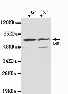
TAB1(N-terminus) Antibody
Purified Mouse Monoclonal Antibody (Mab)
- SPECIFICATION
- CITATIONS
- PROTOCOLS
- BACKGROUND

Application
| WB |
|---|---|
| Primary Accession | Q15750 |
| Reactivity | Human |
| Host | Mouse |
| Clonality | Monoclonal |
| Calculated MW | 55 KDa |
| Gene ID | 10454 |
|---|---|
| Other Names | 2310012M03Rik;3'-Tab1;MAP3K7IP 1;MAP3K7IP1;MGC57664;Mitogen activated protein kinase kinase kinase 7 interacting protein 1;Mitogen-activated protein kinase kinase kinase 7-interacting protein 1;TAB 1;TAB1;TAB1_HUMAN;TAK1 binding protein 1;TAK1-binding protein 1;TGF beta activated kinase 1 binding protein 1;TGF-beta activated kinase 1/MAP3K7 binding protein 1;TGF-beta-activated kinase 1 and MAP3K7-binding protein 1;TGF-beta-activated kinase 1-binding protein 1;Transforming growth factor beta activated kinase binding protein 1. |
| Dilution | WB~~1:1000 |
| Format | Purified mouse monoclonal in buffer containing 0.1M Tris-Glycine (pH 7.4, 150 mM NaCl) with 0.09% (W/V) sodium azide, 50%,glycerol |
| Storage | Store at -20 °C.Stable for 12 months from date of receipt |
| Name | TAB1 |
|---|---|
| Synonyms | MAP3K7IP1 |
| Function | Key adapter protein that plays an essential role in JNK and NF-kappa-B activation and proinflammatory cytokines production in response to stimulation with TLRs and cytokines (PubMed:22307082, PubMed:24403530). Mechanistically, associates with the catalytic domain of MAP3K7/TAK1 to trigger MAP3K7/TAK1 autophosphorylation leading to its full activation (PubMed:10838074, PubMed:25260751, PubMed:37832545). Similarly, associates with MAPK14 and triggers its autophosphorylation and subsequent activation (PubMed:11847341, PubMed:29229647). In turn, MAPK14 phosphorylates TAB1 and inhibits MAP3K7/TAK1 activation in a feedback control mechanism (PubMed:14592977). Also plays a role in recruiting MAPK14 to the TAK1 complex for the phosphorylation of the TAB2 and TAB3 regulatory subunits (PubMed:18021073). |
| Cellular Location | Cytoplasm, cytosol. Endoplasmic reticulum membrane; Peripheral membrane protein; Cytoplasmic side. Note=Recruited to the endoplasmic reticulum following interaction with STING1 |
| Tissue Location | Ubiquitous.. |

Thousands of laboratories across the world have published research that depended on the performance of antibodies from Abcepta to advance their research. Check out links to articles that cite our products in major peer-reviewed journals, organized by research category.
info@abcepta.com, and receive a free "I Love Antibodies" mug.

Provided below are standard protocols that you may find useful for product applications.
Background
May be an important signaling intermediate between TGFB receptors and MAP3K7/TAK1. May play an important role in mammalian embryogenesis.
References
Shibuya H.,et al.Science 272:1179-1182(1996).
Ge B.,et al.J. Biol. Chem. 278:2286-2293(2003).
Dunham I.,et al.Nature 402:489-495(1999).
Mural R.J.,et al.Submitted (JUL-2005) to the EMBL/GenBank/DDBJ databases.
Ninomiya-Tsuji J.,et al.Nature 398:252-256(1999).
If you have used an Abcepta product and would like to share how it has performed, please click on the "Submit Review" button and provide the requested information. Our staff will examine and post your review and contact you if needed.
If you have any additional inquiries please email technical services at tech@abcepta.com.
Ordering Information
Other Products
Shipping Information
