


 Foundational characteristics of cancer include proliferation, angiogenesis, migration, evasion of apoptosis, and cellular immortality. Find key markers for these cellular processes and antibodies to detect them.
 The SUMOplot™ Analysis Program predicts and scores sumoylation sites in your protein. SUMOylation is a post-translational modification involved in various cellular processes, such as nuclear-cytosolic transport, transcriptional regulation, apoptosis, protein stability, response to stress, and progression through the cell cycle.
 The Autophagy Receptor Motif Plotter predicts and scores autophagy receptor binding sites in your protein. Identifying proteins connected to this pathway is critical to understanding the role of autophagy in physiological as well as pathological processes such as development, differentiation, neurodegenerative diseases, stress, infection, and cancer.
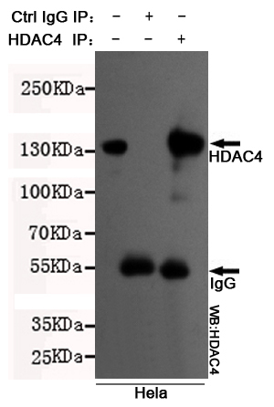
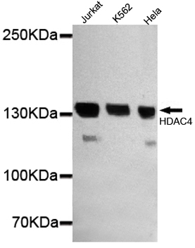
HDAC4 (N-terminus) Antibody
Purified Mouse Monoclonal Antibody (Mab)
- SPECIFICATION
- CITATIONS
- PROTOCOLS
- BACKGROUND

Application
| WB, IP |
|---|---|
| Primary Accession | P56524 |
| Reactivity | Human |
| Host | Mouse |
| Clonality | Monoclonal |
| Isotype | IgG2a |
| Calculated MW | 140 KDa |
| Gene ID | 9759 |
|---|---|
| Other Names | EC 3.5.1.98;HA6116;HD 4;HD4;HDAC 4;HDAC A;HDAC4;HDAC4_HUMAN;HDACA;Histone Deacetylase 4; Histone Deacetylase A;KIAA0288. |
| Dilution | WB~~1:1000 IP~~1:500 |
| Format | Liquid in PBS containing 50% glycerol, 0.5% BSA and 0.02% sodium azide, pH 7.3. |
| Storage | Store at 4°C short term. Aliquot and store at -20°C long term. Avoid freeze/thaw cycles. |
| Name | HDAC4 (HGNC:14063) |
|---|---|
| Synonyms | KIAA0288 |
| Function | Responsible for the deacetylation of lysine residues on the N-terminal part of the core histones (H2A, H2B, H3 and H4). Histone deacetylation gives a tag for epigenetic repression and plays an important role in transcriptional regulation, cell cycle progression and developmental events. Histone deacetylases act via the formation of large multiprotein complexes. Involved in muscle maturation via its interaction with the myocyte enhancer factors such as MEF2A, MEF2C and MEF2D. Involved in the MTA1-mediated epigenetic regulation of ESR1 expression in breast cancer. Deacetylates HSPA1A and HSPA1B at 'Lys-77' leading to their preferential binding to co-chaperone STUB1 (PubMed:27708256). |
| Cellular Location | Nucleus. Cytoplasm. Note=Shuttles between the nucleus and the cytoplasm. Upon muscle cells differentiation, it accumulates in the nuclei of myotubes, suggesting a positive role of nuclear HDAC4 in muscle differentiation. The export to cytoplasm depends on the interaction with a 14-3-3 chaperone protein and is due to its phosphorylation at Ser-246, Ser-467 and Ser-632 by CaMK4 and SIK1. The nuclear localization probably depends on sumoylation Interaction with SIK3 leads to HDAC4 retention in the cytoplasm (By similarity). {ECO:0000250|UniProtKB:Q6NZM9} |
| Tissue Location | Ubiquitous. |

Thousands of laboratories across the world have published research that depended on the performance of antibodies from Abcepta to advance their research. Check out links to articles that cite our products in major peer-reviewed journals, organized by research category.
info@abcepta.com, and receive a free "I Love Antibodies" mug.

Provided below are standard protocols that you may find useful for product applications.
Background
Responsible for the deacetylation of lysine residues on the N-terminal part of the core histones (H2A, H2B, H3 and H4). Histone deacetylation gives a tag for epigenetic repression and plays an important role in transcriptional regulation, cell cycle progression and developmental events. Histone deacetylases act via the formation of large multiprotein complexes. Involved in muscle maturation via its interaction with the myocyte enhancer factors such as MEF2A, MEF2C and MEF2D. Involved in the MTA1-mediated epigenetic regulation of ESR1 expression in breast cancer.
References
Grozinger C.M.,et al.Proc. Natl. Acad. Sci. U.S.A. 96:4868-4873(1999).
Ohara O.,et al.DNA Res. 4:53-59(1997).
Ohara O.,et al.Submitted (DEC-1999) to the EMBL/GenBank/DDBJ databases.
Hillier L.W.,et al.Nature 434:724-731(2005).
Mural R.J.,et al.Submitted (SEP-2005) to the EMBL/GenBank/DDBJ databases.
If you have used an Abcepta product and would like to share how it has performed, please click on the "Submit Review" button and provide the requested information. Our staff will examine and post your review and contact you if needed.
If you have any additional inquiries please email technical services at tech@abcepta.com.
Ordering Information
Other Products
Shipping Information
