


 Foundational characteristics of cancer include proliferation, angiogenesis, migration, evasion of apoptosis, and cellular immortality. Find key markers for these cellular processes and antibodies to detect them.
 The SUMOplot™ Analysis Program predicts and scores sumoylation sites in your protein. SUMOylation is a post-translational modification involved in various cellular processes, such as nuclear-cytosolic transport, transcriptional regulation, apoptosis, protein stability, response to stress, and progression through the cell cycle.
 The Autophagy Receptor Motif Plotter predicts and scores autophagy receptor binding sites in your protein. Identifying proteins connected to this pathway is critical to understanding the role of autophagy in physiological as well as pathological processes such as development, differentiation, neurodegenerative diseases, stress, infection, and cancer.
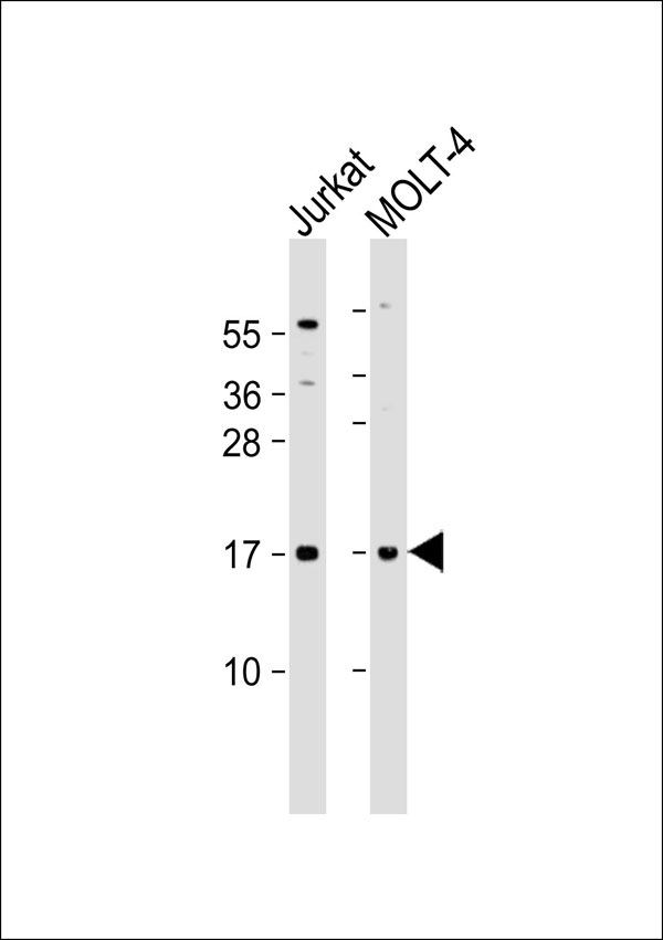
CD3d Antibody
Purified Rabbit Polyclonal Antibody (Pab)
- SPECIFICATION
- CITATIONS
- PROTOCOLS
- BACKGROUND

Application
| WB |
|---|---|
| Primary Accession | P04234 |
| Reactivity | Human |
| Host | Rabbit |
| Clonality | Polyclonal |
| Calculated MW | 19 KDa |
| Antigen Region | 52-101 aa |
| Gene ID | 915 |
|---|---|
| Other Names | T-cell surface glycoprotein CD3 delta chain, T-cell receptor T3 delta chain, CD3d, CD3D, T3D |
| Target/Specificity | KLH-conjugated synthetic peptide encompassing a sequence within the center region of human CD3d. The exact sequence is proprietary. |
| Dilution | WB~~ 1:500 |
| Format | Rabbit IgG in phosphate buffered saline , pH 7.4, 150mM NaCl, 0.09% (W/V) sodium azide and 50% glycerol |
| Storage | Store at -20 °C.Stable for 12 months from date of receipt |
| Name | CD3D |
|---|---|
| Synonyms | T3D |
| Function | Part of the TCR-CD3 complex present on T-lymphocyte cell surface that plays an essential role in adaptive immune response. When antigen presenting cells (APCs) activate T-cell receptor (TCR), TCR- mediated signals are transmitted across the cell membrane by the CD3 chains CD3D, CD3E, CD3G and CD3Z. All CD3 chains contain immunoreceptor tyrosine-based activation motifs (ITAMs) in their cytoplasmic domain. Upon TCR engagement, these motifs become phosphorylated by Src family protein tyrosine kinases LCK and FYN, resulting in the activation of downstream signaling pathways (PubMed:2470098). In addition of this role of signal transduction in T-cell activation, CD3D plays an essential role in thymocyte differentiation. Indeed, participates in correct intracellular TCR-CD3 complex assembly and surface expression. In absence of a functional TCR-CD3 complex, thymocytes are unable to differentiate properly. Interacts with CD4 and CD8 and thus serves to establish a functional link between the TCR and coreceptors CD4 and CD8, which is needed for activation and positive selection of CD4 or CD8 T-cells (PubMed:12215456). |
| Cellular Location | Cell membrane; Single-pass type I membrane protein |
| Tissue Location | CD3D is mostly present on T-lymphocytes with its TCR-CD3 partners. Present also in fetal NK-cells |

Thousands of laboratories across the world have published research that depended on the performance of antibodies from Abcepta to advance their research. Check out links to articles that cite our products in major peer-reviewed journals, organized by research category.
info@abcepta.com, and receive a free "I Love Antibodies" mug.

Provided below are standard protocols that you may find useful for product applications.
Background
The CD3 complex mediates signal transduction.
References
van den Elsen P.,et al.Proc. Natl. Acad. Sci. U.S.A. 83:2944-2948(1986).
van den Elsen P.,et al.Nature 312:413-418(1984).
Tunnacliffe A.,et al.EMBO J. 5:1245-1252(1986).
Jin P.,et al.Genomics 83:566-571(2004).
Taylor T.D.,et al.Nature 440:497-500(2006).
If you have used an Abcepta product and would like to share how it has performed, please click on the "Submit Review" button and provide the requested information. Our staff will examine and post your review and contact you if needed.
If you have any additional inquiries please email technical services at tech@abcepta.com.
Ordering Information
Other Products
Shipping Information
