


 Foundational characteristics of cancer include proliferation, angiogenesis, migration, evasion of apoptosis, and cellular immortality. Find key markers for these cellular processes and antibodies to detect them.
 The SUMOplot™ Analysis Program predicts and scores sumoylation sites in your protein. SUMOylation is a post-translational modification involved in various cellular processes, such as nuclear-cytosolic transport, transcriptional regulation, apoptosis, protein stability, response to stress, and progression through the cell cycle.
 The Autophagy Receptor Motif Plotter predicts and scores autophagy receptor binding sites in your protein. Identifying proteins connected to this pathway is critical to understanding the role of autophagy in physiological as well as pathological processes such as development, differentiation, neurodegenerative diseases, stress, infection, and cancer.
RhoG Antibody
Purified Rabbit Polyclonal Antibody (Pab)
- SPECIFICATION
- CITATIONS
- PROTOCOLS
- BACKGROUND

Application
| WB |
|---|---|
| Primary Accession | P84095 |
| Reactivity | Human, Mouse |
| Host | Rabbit |
| Clonality | Polyclonal |
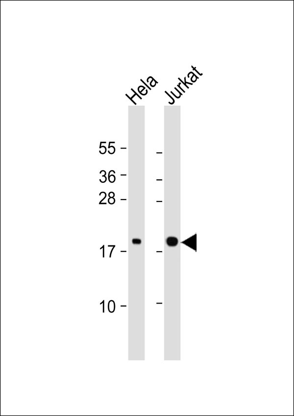
| Calculated MW | 21 KDa |
| Antigen Region | 97-146 aa |
| Gene ID | 391 |
|---|---|
| Other Names | Rho-related GTP-binding protein RhoG, RHOG, ARHG |
| Target/Specificity | KLH-conjugated synthetic peptide encompassing a sequence within the center region of human RhoG. The exact sequence is proprietary. |
| Dilution | WB~~ 1:1000 |
| Format | Rabbit IgG in phosphate buffered saline , pH 7.4, 150mM NaCl, 0.09% (W/V) sodium azide and 50% glycerol |
| Storage | Store at -20 °C.Stable for 12 months from date of receipt |
| Name | RHOG |
|---|---|
| Synonyms | ARHG |
| Function | Plays a role in immunological synaptic F-actin density and architecture organization (PubMed:33513601). Regulates actin reorganization in lymphocytes, possibly through the modulation of Rac1 activity (PubMed:33513601). Required for the formation of membrane ruffles during macropinocytosis (PubMed:15133129). Plays a role in cell migration and is required for the formation of cup-like structures during trans-endothelial migration of leukocytes (PubMed:17875742). Binds phospholipids in an activation-dependent manner; thereby acting as an anchor for other proteins to the plasma membrane (PM) (PubMed:33513601). Plays a role in exocytosis of cytotoxic granules (CG) by lymphocytes/Component of the exocytosis machinery in natural killer (NK) and CD8+ T cells (PubMed:33513601). Promotes the docking of cytotoxic granules (CG) to the plasma membrane through the interaction with UNC13D (PubMed:33513601). Involved in the cytotoxic activity of lymphocytes/primary CD8+ T cells (PubMed:33513601). |
| Cellular Location | Cell membrane; Lipid-anchor; Cytoplasmic side |

Thousands of laboratories across the world have published research that depended on the performance of antibodies from Abcepta to advance their research. Check out links to articles that cite our products in major peer-reviewed journals, organized by research category.
info@abcepta.com, and receive a free "I Love Antibodies" mug.

Provided below are standard protocols that you may find useful for product applications.
Background
Required for the formation of membrane ruffles during macropinocytosis. Plays a role in cell migration and is required for the formation of cup-like structures during trans-endothelial migration of leukocytes. In case of Salmonella enterica infection, activated by SopB and ARHGEF26/SGEF, which induces cytoskeleton rearrangements and promotes bacterial entry.
References
Vincent S.,et al.Mol. Cell. Biol. 12:3138-3148(1992).
Miki T.,et al.Nature 362:462-465(1993).
Puhl H.L. III,et al.Submitted (FEB-2004) to the EMBL/GenBank/DDBJ databases.
Ellerbroek S.M.,et al.Mol. Biol. Cell 15:3309-3319(2004).
Patel J.C.,et al.J. Cell Biol. 175:453-463(2006).
If you have used an Abcepta product and would like to share how it has performed, please click on the "Submit Review" button and provide the requested information. Our staff will examine and post your review and contact you if needed.
If you have any additional inquiries please email technical services at tech@abcepta.com.
Ordering Information
Shipping Information
