


 Foundational characteristics of cancer include proliferation, angiogenesis, migration, evasion of apoptosis, and cellular immortality. Find key markers for these cellular processes and antibodies to detect them.
 The SUMOplot™ Analysis Program predicts and scores sumoylation sites in your protein. SUMOylation is a post-translational modification involved in various cellular processes, such as nuclear-cytosolic transport, transcriptional regulation, apoptosis, protein stability, response to stress, and progression through the cell cycle.
 The Autophagy Receptor Motif Plotter predicts and scores autophagy receptor binding sites in your protein. Identifying proteins connected to this pathway is critical to understanding the role of autophagy in physiological as well as pathological processes such as development, differentiation, neurodegenerative diseases, stress, infection, and cancer.
Anti-DGAT2 Antibody
- SPECIFICATION
- CITATIONS
- PROTOCOLS
- BACKGROUND

Application
| WB |
|---|---|
| Primary Accession | Q96PD7 |
| Reactivity | Human, Mouse, Rat |
| Host | Rabbit |
| Clonality | Polyclonal |
| Calculated MW | 43831 Da |
| Gene ID | 84649 |
|---|---|
| Other Names | Diacylglycerol O-acyltransferase 2; Acyl-CoA retinol O-fatty-acyltransferase; ARAT; Retinol O-fatty-acyltransferase; Diglyceride acyltransferase 2 |
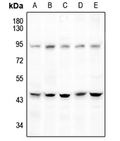
| Target/Specificity | Recognizes endogenous levels of DGAT2 protein. |
| Dilution | WB~~1/500 - 1/1000 |
| Format | Liquid in 0.42% Potassium phosphate, 0.87% Sodium chloride, pH 7.3, 30% glycerol, and 0.09% (W/V) sodium azide. |
| Storage | Store at -20 °C.Stable for 12 months from date of receipt |
| Name | DGAT2 (HGNC:16940) |
|---|---|
| Function | Essential acyltransferase that catalyzes the terminal and only committed step in triacylglycerol synthesis by using diacylglycerol and fatty acyl CoA as substrates. Required for synthesis and storage of intracellular triglycerides (PubMed:27184406). Probably plays a central role in cytosolic lipid accumulation. In liver, is primarily responsible for incorporating endogenously synthesized fatty acids into triglycerides (By similarity). Also functions as an acyl-CoA retinol acyltransferase (ARAT) (By similarity). Also able to use 1- monoalkylglycerol (1-MAkG) as an acyl acceptor for the synthesis of monoalkyl-monoacylglycerol (MAMAG) (PubMed:28420705). |
| Cellular Location | Endoplasmic reticulum membrane; Multi-pass membrane protein. Lipid droplet. Cytoplasm, perinuclear region |
| Tissue Location | Predominantly expressed in liver and white adipose tissue. Expressed at lower level in mammary gland, testis and peripheral blood leukocytes. Expressed in sebaceous glands of normal skin but decreased psoriatic skin. |
Research Areas
Citations (0)

Thousands of laboratories across the world have published research that depended on the performance of antibodies from Abcepta to advance their research. Check out links to articles that cite our products in major peer-reviewed journals, organized by research category.
Submit your citation using an Abcepta antibody to
info@abcepta.com, and receive a free "I Love Antibodies" mug.
info@abcepta.com, and receive a free "I Love Antibodies" mug.

Application Protocols
Provided below are standard protocols that you may find useful for product applications.
Background
Rabbit polyclonal antibody to DGAT2
Abcepta welcomes feedback from its customers.
If you have used an Abcepta product and would like to share how it has performed, please click on the "Submit Review" button and provide the requested information. Our staff will examine and post your review and contact you if needed.
If you have any additional inquiries please email technical services at tech@abcepta.com.
$ 385.00
Cat# AP53746
Ordering Information
United States
AlbaniaAustraliaAustriaBelgiumBosnia & HerzegovinaBrazilBulgariaCanadaCentral AmericaChinaCroatiaCyprusCzech RepublicDenmarkEstoniaFinlandFranceGermanyGreeceHong KongHungaryIcelandIndiaIndonesiaIrelandIsraelItalyJapanLatviaLithuaniaLuxembourgMacedoniaMalaysiaMaltaMexicoNetherlandsNew ZealandNorwayPakistanPolandPortugalRomaniaSerbiaSingaporeSlovakiaSloveniaSouth AfricaSouth KoreaSpainSwedenSwitzerlandTaiwanTurkeyUnited KingdomUnited StatesVietnamWorldwideOthers
USA Headquarters
(888) 735-7227 / (858) 622-0099 or (858) 875-1900
Other Products
Shipping Information
Domestic orders (in stock items)
Shipped out the same day. Orders placed after 1 PM (PST) will ship out the next business day.
International orders
Contact your local distributors
