


 Foundational characteristics of cancer include proliferation, angiogenesis, migration, evasion of apoptosis, and cellular immortality. Find key markers for these cellular processes and antibodies to detect them.
 The SUMOplot™ Analysis Program predicts and scores sumoylation sites in your protein. SUMOylation is a post-translational modification involved in various cellular processes, such as nuclear-cytosolic transport, transcriptional regulation, apoptosis, protein stability, response to stress, and progression through the cell cycle.
 The Autophagy Receptor Motif Plotter predicts and scores autophagy receptor binding sites in your protein. Identifying proteins connected to this pathway is critical to understanding the role of autophagy in physiological as well as pathological processes such as development, differentiation, neurodegenerative diseases, stress, infection, and cancer.
> home > Products > Primary Antibodies > Antibody Collections > Urinary bladder negative > Anti-MYO1D Antibody
Anti-MYO1D Antibody
- SPECIFICATION
- CITATIONS
- PROTOCOLS
- BACKGROUND

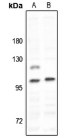
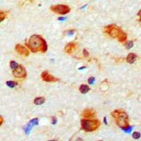
Application
| WB, IHC |
|---|---|
| Primary Accession | O94832 |
| Reactivity | Human, Mouse, Rat |
| Host | Rabbit |
| Clonality | Polyclonal |
| Calculated MW | 116202 Da |
| Gene ID | 4642 |
|---|---|
| Other Names | KIAA0727; Unconventional myosin-Id |
| Target/Specificity | KLH-conjugated synthetic peptide encompassing a sequence within the C-term region of human MYO1D. The exact sequence is proprietary. |
| Dilution | WB~~1/500 - 1/1000 IHC~~1:100~500 |
| Format | Liquid in 0.42% Potassium phosphate, 0.87% Sodium chloride, pH 7.3, 30% glycerol, and 0.09% (W/V) sodium azide. |
| Storage | Store at -20 °C.Stable for 12 months from date of receipt |
| Name | MYO1D |
|---|---|
| Synonyms | KIAA0727 |
| Function | Unconventional myosin that functions as actin-based motor protein with ATPase activity (By similarity). Plays a role in endosomal protein trafficking, and especially in the transfer of cargo proteins from early to recycling endosomes (By similarity). Required for normal planar cell polarity in ciliated tracheal cells, for normal rotational polarity of cilia, and for coordinated, unidirectional ciliary movement in the trachea. Required for normal, polarized cilia organization in brain ependymal epithelial cells (By similarity). |
| Cellular Location | Cytoplasm {ECO:0000250|UniProtKB:Q63357}. Perikaryon {ECO:0000250|UniProtKB:Q63357}. Cell projection, dendrite {ECO:0000250|UniProtKB:Q63357}. Early endosome {ECO:0000250|UniProtKB:F1PRN2}. Cytoplasm, cell cortex {ECO:0000250|UniProtKB:Q63357}. Note=Colocalizes with the actin cytoskeleton in the cell cortex close to the apical cell membrane Colocalizes with cytoplasmic puncta that are reminiscent of transport vesicles. {ECO:0000250|UniProtKB:Q63357} |
| Tissue Location | Expressed in many tissues. Highest levels in brain, followed by lung and ovary; expression is lowest in spleen |
Research Areas
Citations (0)

Thousands of laboratories across the world have published research that depended on the performance of antibodies from Abcepta to advance their research. Check out links to articles that cite our products in major peer-reviewed journals, organized by research category.
Submit your citation using an Abcepta antibody to
info@abcepta.com, and receive a free "I Love Antibodies" mug.
info@abcepta.com, and receive a free "I Love Antibodies" mug.

Application Protocols
Provided below are standard protocols that you may find useful for product applications.
Background
Rabbit polyclonal antibody to MYO1D
Abcepta welcomes feedback from its customers.
If you have used an Abcepta product and would like to share how it has performed, please click on the "Submit Review" button and provide the requested information. Our staff will examine and post your review and contact you if needed.
If you have any additional inquiries please email technical services at tech@abcepta.com.
$ 385.00
Cat# AP53856
Ordering Information
United States
AlbaniaAustraliaAustriaBelgiumBosnia & HerzegovinaBrazilBulgariaCanadaCentral AmericaChinaCroatiaCyprusCzech RepublicDenmarkEstoniaFinlandFranceGermanyGreeceHong KongHungaryIcelandIndiaIndonesiaIrelandIsraelItalyJapanLatviaLithuaniaLuxembourgMacedoniaMalaysiaMaltaMexicoNetherlandsNew ZealandNorwayPakistanPolandPortugalRomaniaSerbiaSingaporeSlovakiaSloveniaSouth AfricaSouth KoreaSpainSwedenSwitzerlandTaiwanTurkeyUnited KingdomUnited StatesVietnamWorldwideOthers
USA Headquarters
(888) 735-7227 / (858) 622-0099 or (858) 875-1900
Other Products
Shipping Information
Domestic orders (in stock items)
Shipped out the same day. Orders placed after 1 PM (PST) will ship out the next business day.
International orders
Contact your local distributors
