


 Foundational characteristics of cancer include proliferation, angiogenesis, migration, evasion of apoptosis, and cellular immortality. Find key markers for these cellular processes and antibodies to detect them.
 The SUMOplot™ Analysis Program predicts and scores sumoylation sites in your protein. SUMOylation is a post-translational modification involved in various cellular processes, such as nuclear-cytosolic transport, transcriptional regulation, apoptosis, protein stability, response to stress, and progression through the cell cycle.
 The Autophagy Receptor Motif Plotter predicts and scores autophagy receptor binding sites in your protein. Identifying proteins connected to this pathway is critical to understanding the role of autophagy in physiological as well as pathological processes such as development, differentiation, neurodegenerative diseases, stress, infection, and cancer.
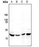
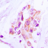
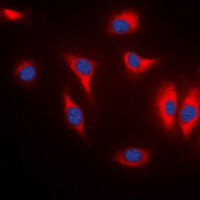
Anti-MARK Antibody
- SPECIFICATION
- CITATIONS
- PROTOCOLS
- BACKGROUND

Application
| WB, IF, IHC |
|---|---|
| Primary Accession | Q9P0L2 |
| Other Accession | Q7KZI7, P27448, Q96L34 |
| Reactivity | Human, Mouse, Rat |
| Host | Rabbit |
| Clonality | Polyclonal |
| Calculated MW | 89003 Da |
| Gene ID | 4139 |
|---|---|
| Other Names | MARK1; KIAA1477; MARK; Serine/threonine-protein kinase MARK1; MAP/microtubule affinity-regulating kinase 1; PAR1 homolog c; Par-1c; Par1c; MARK2; EMK1; Serine/threonine-protein kinase MARK2; ELKL motif kinase 1; EMK-1; MAP/microtubule affinity-regulating kinase 2; PAR1 homolog; PAR1 homolog b; Par-1b; Par1b; MARK3; CTAK1; EMK2; MAP/microtubule affinity-regulating kinase 3; C-TAK1; cTAK1; Cdc25C-associated protein kinase 1; ELKL motif kinase 2; EMK-2; Protein kinase STK10; Ser/Thr protein kinase PAR-1; Par-1a; Serine/threonine-protein kinase p78; MARK4; KIAA1860; MARKL1; MAP/microtubule affinity-regulating kinase 4; MAP/microtubule affinity-regulating kinase-like 1 |
| Target/Specificity | KLH-conjugated synthetic peptide encompassing a sequence within the center region of human MARK. The exact sequence is proprietary. |
| Dilution | WB~~1/500 - 1/1000 IF~~1/50 - 1/200 IHC~~1:100~500 |
| Format | Liquid in 0.42% Potassium phosphate, 0.87% Sodium chloride, pH 7.3, 30% glycerol, and 0.09% (W/V) sodium azide. |
| Storage | Store at -20 °C.Stable for 12 months from date of receipt |
| Name | MARK1 (HGNC:6896) |
|---|---|
| Function | Serine/threonine-protein kinase (PubMed:23666762). Involved in cell polarity and microtubule dynamics regulation. Phosphorylates DCX, MAP2 and MAP4. Phosphorylates the microtubule-associated protein MAPT/TAU (PubMed:23666762). Involved in cell polarity by phosphorylating the microtubule-associated proteins MAP2, MAP4 and MAPT/TAU at KXGS motifs, causing detachment from microtubules, and their disassembly. Involved in the regulation of neuronal migration through its dual activities in regulating cellular polarity and microtubule dynamics, possibly by phosphorylating and regulating DCX. Also acts as a positive regulator of the Wnt signaling pathway, probably by mediating phosphorylation of dishevelled proteins (DVL1, DVL2 and/or DVL3). |
| Cellular Location | Cell membrane; Peripheral membrane protein. Cytoplasm, cytoskeleton. Cytoplasm Cell projection, dendrite. Note=Appears to localize to an intracellular network. |
| Tissue Location | Highly expressed in heart, skeletal muscle, brain, fetal brain and fetal kidney. |
Research Areas
Citations (0)

Thousands of laboratories across the world have published research that depended on the performance of antibodies from Abcepta to advance their research. Check out links to articles that cite our products in major peer-reviewed journals, organized by research category.
Submit your citation using an Abcepta antibody to
info@abcepta.com, and receive a free "I Love Antibodies" mug.
info@abcepta.com, and receive a free "I Love Antibodies" mug.

Application Protocols
Provided below are standard protocols that you may find useful for product applications.
Background
Rabbit polyclonal antibody to MARK
Abcepta welcomes feedback from its customers.
If you have used an Abcepta product and would like to share how it has performed, please click on the "Submit Review" button and provide the requested information. Our staff will examine and post your review and contact you if needed.
If you have any additional inquiries please email technical services at tech@abcepta.com.
$ 385.00
Cat# AP53905
Ordering Information
United States
AlbaniaAustraliaAustriaBelgiumBosnia & HerzegovinaBrazilBulgariaCanadaCentral AmericaChinaCroatiaCyprusCzech RepublicDenmarkEstoniaFinlandFranceGermanyGreeceHong KongHungaryIcelandIndiaIndonesiaIrelandIsraelItalyJapanLatviaLithuaniaLuxembourgMacedoniaMalaysiaMaltaMexicoNetherlandsNew ZealandNorwayPakistanPolandPortugalRomaniaSerbiaSingaporeSlovakiaSloveniaSouth AfricaSouth KoreaSpainSwedenSwitzerlandTaiwanTurkeyUnited KingdomUnited StatesVietnamWorldwideOthers
USA Headquarters
(888) 735-7227 / (858) 622-0099 or (858) 875-1900
Other Products
Shipping Information
Domestic orders (in stock items)
Shipped out the same day. Orders placed after 1 PM (PST) will ship out the next business day.
International orders
Contact your local distributors
