


 Foundational characteristics of cancer include proliferation, angiogenesis, migration, evasion of apoptosis, and cellular immortality. Find key markers for these cellular processes and antibodies to detect them.
 The SUMOplot™ Analysis Program predicts and scores sumoylation sites in your protein. SUMOylation is a post-translational modification involved in various cellular processes, such as nuclear-cytosolic transport, transcriptional regulation, apoptosis, protein stability, response to stress, and progression through the cell cycle.
 The Autophagy Receptor Motif Plotter predicts and scores autophagy receptor binding sites in your protein. Identifying proteins connected to this pathway is critical to understanding the role of autophagy in physiological as well as pathological processes such as development, differentiation, neurodegenerative diseases, stress, infection, and cancer.
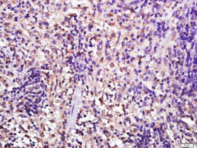
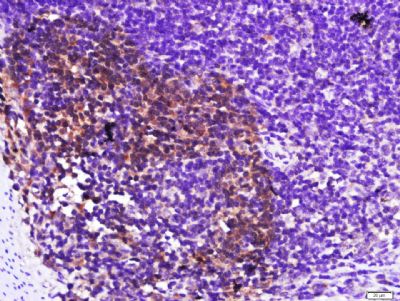
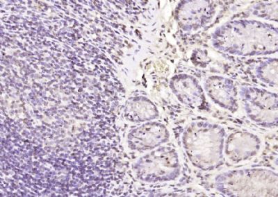
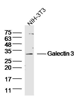
Galectin 3 Polyclonal Antibody
Purified Rabbit Polyclonal Antibody (Pab)
- SPECIFICATION
- CITATIONS
- PROTOCOLS
- BACKGROUND

Application
| IHC-P, IHC-F, IF, ICC, E |
|---|---|
| Primary Accession | P16110 |
| Reactivity | Rat |
| Host | Rabbit |
| Clonality | Polyclonal |
| Calculated MW | 29 KDa |
| Physical State | Liquid |
| Immunogen | KLH conjugated synthetic peptide derived from mouse Galectin-3 |
| Epitope Specificity | 181-262/262 |
| Isotype | IgG |
| Purity | affinity purified by Protein A |
| Buffer | 0.01M TBS (pH7.4) with 1% BSA, 0.02% Proclin300 and 50% Glycerol. |
| SUBCELLULAR LOCATION | Cytoplasm. Nucleus. Secreted. Note=Secreted by a non-classical secretory pathway and associates with the cell surface. |
| SIMILARITY | Contains 1 galectin domain. |
| SUBUNIT | Probably forms homo- or heterodimers. Interacts with DMBT1 (By similarity). Forms a complex with the ITGA3, ITGB1 and CSPG4. Interacts with LGALS3BP, LYPD3, CYHR1 and UACA. |
| Important Note | This product as supplied is intended for research use only, not for use in human, therapeutic or diagnostic applications. |
| Background Descriptions | This gene encodes a member of thegalectinfamily of carbohydrate binding proteins. Members of this protein family have an affinity for beta-galactosides. The encoded protein is characterized by an N-terminal proline-rich tandem repeat domain and a single C-terminal carbohydrate recognition domain. This protein can self-associate through the N-terminal domain allowing it to bind to multivalent saccharide ligands. This protein localizes to the extracellular matrix, the cytoplasm and the nucleus. This protein plays a role in numerous cellular functions including apoptosis, innate immunity, cell adhesion and T-cell regulation. The protein exhibits antimicrobial activity against bacteria and fungi. Alternate splicing results in multiple transcript variants.[provided by RefSeq, Oct 2014] |
| Other Names | Galectin-3, Gal-3, 35 kDa lectin, Carbohydrate-binding protein 35, CBP 35, Galactose-specific lectin 3, IgE-binding protein, L-34 galactoside-binding lectin, Laminin-binding protein, Lectin L-29, Mac-2 antigen, Lgals3 |
|---|---|
| Target/Specificity | A major expression is found in the colonic epithelium. It is also abundant in the activated macrophages. |
| Dilution | IHC-P=1:100-500,IHC-F=1:100-500,ICC=1:100-500,IF=1:100-500,ELISA=1:5000-10000 |
| Storage | Store at -20 ℃ for one year. Avoid repeated freeze/thaw cycles. When reconstituted in sterile pH 7.4 0.01M PBS or diluent of antibody the antibody is stable for at least two weeks at 2-4 ℃. |
| Name | Lgals3 |
|---|---|
| Function | Galactose-specific lectin which binds IgE. May mediate with the alpha-3, beta-1 integrin the stimulation by CSPG4 of endothelial cells migration (PubMed:15181153). Together with DMBT1, required for terminal differentiation of columnar epithelial cells during early embryogenesis. In the nucleus: acts as a pre-mRNA splicing factor. Involved in acute inflammatory responses including neutrophil activation and adhesion, chemoattraction of monocytes macrophages, opsonization of apoptotic neutrophils, and activation of mast cells. Together with TRIM16, coordinates the recognition of membrane damage with mobilization of the core autophagy regulators ATG16L1 and BECN1 in response to damaged endomembranes (By similarity). When secreted, interacts with NK cell-activating receptor NCR3/NKp30 acting as an inhibitory ligand which antagonizes NK cell attack (By similarity). |
| Cellular Location | Cytoplasm {ECO:0000250|UniProtKB:P17931}. Nucleus {ECO:0000250|UniProtKB:P17931}. Secreted {ECO:0000250|UniProtKB:P17931}. Note=Secreted by a non-classical secretory pathway and associates with the cell surface. Can be secreted; the secretion is dependent on protein unfolding and facilitated by the cargo receptor TMED10; it results in protein translocation from the cytoplasm into the ERGIC (endoplasmic reticulum- Golgi intermediate compartment) followed by vesicle entry and secretion. {ECO:0000250|UniProtKB:P17931} |
| Tissue Location | The highest levels are found in activated macrophages |
Research Areas
Citations (0)

Thousands of laboratories across the world have published research that depended on the performance of antibodies from Abcepta to advance their research. Check out links to articles that cite our products in major peer-reviewed journals, organized by research category.
Submit your citation using an Abcepta antibody to
info@abcepta.com, and receive a free "I Love Antibodies" mug.
info@abcepta.com, and receive a free "I Love Antibodies" mug.

Application Protocols
Provided below are standard protocols that you may find useful for product applications.
Abcepta welcomes feedback from its customers.
If you have used an Abcepta product and would like to share how it has performed, please click on the "Submit Review" button and provide the requested information. Our staff will examine and post your review and contact you if needed.
If you have any additional inquiries please email technical services at tech@abcepta.com.
$ 385.00
Cat# AP54254
Ordering Information
United States
AlbaniaAustraliaAustriaBelgiumBosnia & HerzegovinaBrazilBulgariaCanadaCentral AmericaChinaCroatiaCyprusCzech RepublicDenmarkEstoniaFinlandFranceGermanyGreeceHong KongHungaryIcelandIndiaIndonesiaIrelandIsraelItalyJapanLatviaLithuaniaLuxembourgMacedoniaMalaysiaMaltaMexicoNetherlandsNew ZealandNorwayPakistanPolandPortugalRomaniaSerbiaSingaporeSlovakiaSloveniaSouth AfricaSouth KoreaSpainSwedenSwitzerlandTaiwanTurkeyUnited KingdomUnited StatesVietnamWorldwideOthers
USA Headquarters
(888) 735-7227 / (858) 622-0099 or (858) 875-1900
Other Products
Shipping Information
Domestic orders (in stock items)
Shipped out the same day. Orders placed after 1 PM (PST) will ship out the next business day.
International orders
Contact your local distributors
