NLGN2 Polyclonal Antibody
Purified Rabbit Polyclonal Antibody (Pab)
- SPECIFICATION
- CITATIONS
- PROTOCOLS
- BACKGROUND

Application
| IHC-P, IHC-F, IF, ICC, E |
|---|---|
| Primary Accession | Q8NFZ4 |
| Reactivity | Rat, Pig, Dog, Bovine |
| Host | Rabbit |
| Clonality | Polyclonal |
| Calculated MW | 90 KDa |
| Physical State | Liquid |
| Immunogen | KLH conjugated synthetic peptide derived from human NLGN1 |
| Epitope Specificity | 251-350/835 |
| Isotype | IgG |
| Purity | affinity purified by Protein A |
| Buffer | 0.01M TBS (pH7.4) with 1% BSA, 0.02% Proclin300 and 50% Glycerol. |
| SUBCELLULAR LOCATION | Cell membrane; Single-pass type I membrane protein. Cell junction, synapse, postsynaptic cell membrane. Cell junction, synapse, presynaptic cell membrane. Note=Detected at postsynaptic membranes in brain. Detected at dendritic spines in cultured neurons. Colocalizes with GPHN and ARHGEF9 at neuronal cell membranes (By similarity). Localized at presynaptic membranes in retina. Colocalizes with GABRG2 at inhibitory synapses in the retina. |
| SIMILARITY | Belongs to the type-B carboxylesterase/lipase family. |
| SUBUNIT | Interacts with NRXN1, NRXN2 and NRXN3. Interacts (via its C-terminus) with DLG4/PSD-95 (via PDZ domain 3). Interacts with INADL. Interacts with GPHN. |
| Important Note | This product as supplied is intended for research use only, not for use in human, therapeutic or diagnostic applications. |
| Background Descriptions | Neuroligins are a family of plasma membrane proteins that possess an N-terminal hydrophobic domain, a large esterase homology domain, a single transmembrane region, a short cytoplasmic domain, and an EF-hand binding domain (1,2). Members of the neuroligin family include Neuroligin 1, Neuroligin 2 and Neuroligin 3. Neuroligins are expressed in excitatory neuronal synaptic clefts. Neuroligins play a role in the formation and remodeling of CNS synapses by binding to b-neurexins, a family of neuronal cell surface proteins. Neuroexin 1b binds to the EF-hand domain of Neuroligin 1 and requires calcium ion. Neuroligins also bind to PSD-95, which may recruit ion channels and neurotransmitter receptors to the synapses. |
| Gene ID | 57555 |
|---|---|
| Other Names | Neuroligin-2, NLGN2, KIAA1366 |
| Target/Specificity | Expressed in the blood vessel walls. Detected in colon, brian and pancreas islets of Langerhans (at protein level). Detected in brain, and at lower levels in pancreas islet beta cells. |
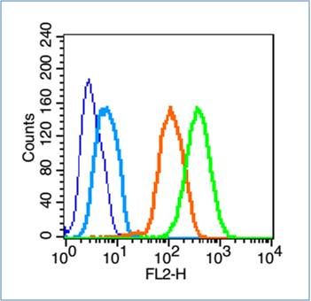
| Dilution | IHC-P=1:100-500,IHC-F=1:100-500,ICC=1:100-500,IF=1:100-500,Flow-Cyt=1 µg/Test,ELISA=1:5000-10000 |
| Format | 0.01M TBS(pH7.4), 0.09% (W/V) sodium azide and 50% Glyce |
| Storage | Store at -20 ℃ for one year. Avoid repeated freeze/thaw cycles. When reconstituted in sterile pH 7.4 0.01M PBS or diluent of antibody the antibody is stable for at least two weeks at 2-4 ℃. |
| Name | NLGN2 |
|---|---|
| Synonyms | KIAA1366 |
| Function | Transmembrane scaffolding protein involved in cell-cell interactions via its interactions with neurexin family members. Mediates cell-cell interactions both in neurons and in other types of cells, such as Langerhans beta cells. Plays a role in synapse function and synaptic signal transmission, especially via gamma-aminobutyric acid receptors (GABA(A) receptors). Functions by recruiting and clustering synaptic proteins. Promotes clustering of postsynaptic GABRG2 and GPHN. Promotes clustering of postsynaptic LHFPL4 (By similarity). Modulates signaling by inhibitory synapses, and thereby plays a role in controlling the ratio of signaling by excitatory and inhibitory synapses and information processing. Required for normal signal amplitude from inhibitory synapses, but is not essential for normal signal frequency. May promote the initial formation of synapses, but is not essential for this. In vitro, triggers the de novo formation of presynaptic structures. Mediates cell-cell interactions between Langerhans beta cells and modulates insulin secretion (By similarity). |
| Cellular Location | Cell membrane; Single-pass type I membrane protein. Postsynaptic cell membrane. Presynaptic cell membrane. Note=Detected at postsynaptic membranes in brain. Detected at dendritic spines in cultured neurons. Colocalizes with GPHN and ARHGEF9 at neuronal cell membranes (By similarity). Localized at presynaptic membranes in retina. Colocalizes with GABRG2 at inhibitory synapses in the retina (By similarity). |
| Tissue Location | Expressed in the blood vessel walls. Detected in colon, brain and pancreas islets of Langerhans (at protein level) Detected in brain, and at lower levels in pancreas islet beta cells |

Thousands of laboratories across the world have published research that depended on the performance of antibodies from Abcepta to advance their research. Check out links to articles that cite our products in major peer-reviewed journals, organized by research category.
info@abcepta.com, and receive a free "I Love Antibodies" mug.
Provided below are standard protocols that you may find useful for product applications.
If you have used an Abcepta product and would like to share how it has performed, please click on the "Submit Review" button and provide the requested information. Our staff will examine and post your review and contact you if needed.
If you have any additional inquiries please email technical services at tech@abcepta.com.


 Foundational characteristics of cancer include proliferation, angiogenesis, migration, evasion of apoptosis, and cellular immortality. Find key markers for these cellular processes and antibodies to detect them.
 The SUMOplot™ Analysis Program predicts and scores sumoylation sites in your protein. SUMOylation is a post-translational modification involved in various cellular processes, such as nuclear-cytosolic transport, transcriptional regulation, apoptosis, protein stability, response to stress, and progression through the cell cycle.
 The Autophagy Receptor Motif Plotter predicts and scores autophagy receptor binding sites in your protein. Identifying proteins connected to this pathway is critical to understanding the role of autophagy in physiological as well as pathological processes such as development, differentiation, neurodegenerative diseases, stress, infection, and cancer.


