


 Foundational characteristics of cancer include proliferation, angiogenesis, migration, evasion of apoptosis, and cellular immortality. Find key markers for these cellular processes and antibodies to detect them.
 The SUMOplot™ Analysis Program predicts and scores sumoylation sites in your protein. SUMOylation is a post-translational modification involved in various cellular processes, such as nuclear-cytosolic transport, transcriptional regulation, apoptosis, protein stability, response to stress, and progression through the cell cycle.
 The Autophagy Receptor Motif Plotter predicts and scores autophagy receptor binding sites in your protein. Identifying proteins connected to this pathway is critical to understanding the role of autophagy in physiological as well as pathological processes such as development, differentiation, neurodegenerative diseases, stress, infection, and cancer.
PRPH2 Polyclonal Antibody
Purified Rabbit Polyclonal Antibody (Pab)
- SPECIFICATION
- CITATIONS
- PROTOCOLS
- BACKGROUND

Application
| WB, IHC-P, IHC-F, IF, ICC, E |
|---|---|
| Primary Accession | P23942 |
| Reactivity | Rat, Dog, Bovine |
| Host | Rabbit |
| Clonality | Polyclonal |
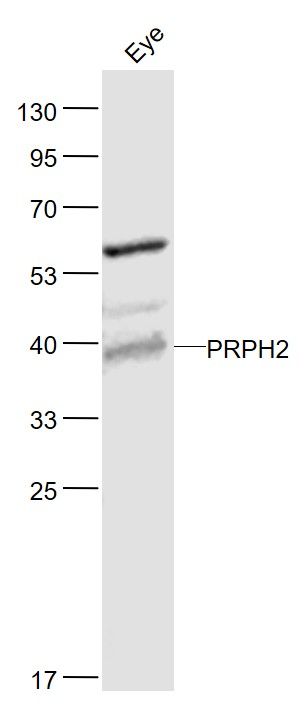
| Calculated MW | 39 KDa |
| Physical State | Liquid |
| Immunogen | KLH conjugated synthetic peptide derived from human PRPH2 |
| Epitope Specificity | 131-230/346 |
| Isotype | IgG |
| Purity | affinity purified by Protein A |
| Buffer | 0.01M TBS (pH7.4) with 1% BSA, 0.02% Proclin300 and 50% Glycerol. |
| SUBCELLULAR LOCATION | Membrane; Multi-pass membrane protein. |
| SIMILARITY | Belongs to the PRPH2/ROM1 family. |
| SUBUNIT | Homodimer; disulfide-linked. Probably forms a complex with a ROM1 homodimer. Other proteins could associate with this complex in rods. Interacts with MREG. |
| DISEASE | Defects in PRPH2 are the cause of retinitis pigmentosa type 7 (RP7). RP leads to degeneration of retinal photoreceptor cells. Patients typically have night vision blindness and loss of midperipheral visual field. As their condition progresses, they lose their far peripheral visual field and eventually central vision as well. Defects in PRPH2 are a cause of retinitis punctata albescens. Defects in PRPH2 are a cause of adult-onset vitelliform macular dystrophy (AVMD). AVMD is a rare autosomal dominant disorder with incomplete penetrance and highly variable expression. Patients usually become symptomatic in the fourth or fifth decade of life with a protracted disease of decreased visual acuity. Defects in PRPH2 are a cause of patterned dystrophy of retinal pigment epithelium (PDREP). Patterned dystrophies of the retinal pigment epithelium (RPE) refer to a heterogeneous group of macular disorders. Three main types of PDREP have been described: reticular (fishnet-like) dystrophy, macroreticular (spider-shaped) dystrophy and butterfly-shaped pigment dystrophy. Defects in PRPH2 are a cause of choroidal dystrophy central areolar type 2 (CACD2). It is a disorder which affects the posterior pole of the eye, and early lesions consist of a non-specific area of granular hyperpigmentation at the fovea. The characteristic sign of the disorder, a zone of atrophy that develops in the macula of the eye and involves the retinal pigment epithelium and the choriocapillaris, occurs several decades after onset. |
| Important Note | This product as supplied is intended for research use only, not for use in human, therapeutic or diagnostic applications. |
| Background Descriptions | May function as an adhesion molecule involved in stabilization and compaction of outer segment disks or in the maintenance of the curvature of the rim. It is essential for disk morphogenesis. |
| Gene ID | 5961 |
|---|---|
| Other Names | Peripherin-2, Retinal degeneration slow protein, Tetraspanin-22, Tspan-22, PRPH2, PRPH, RDS, TSPAN22 |
| Target/Specificity | Retina (photoreceptor). In rim region of ROS (rod outer segment) disks. |
| Dilution | WB=1:500-2000,IHC-P=1:100-500,IHC-F=1:100-500,ICC=1:100-500,IF=1:100-500,ELISA=1:5000-10000 |
| Storage | Store at -20 ℃ for one year. Avoid repeated freeze/thaw cycles. When reconstituted in sterile pH 7.4 0.01M PBS or diluent of antibody the antibody is stable for at least two weeks at 2-4 ℃. |
| Name | PRPH2 |
|---|---|
| Synonyms | PRPH, RDS, TSPAN22 |
| Function | Essential for retina photoreceptor outer segment disk morphogenesis, may also play a role with ROM1 in the maintenance of outer segment disk structure (By similarity). Required for the maintenance of retinal outer nuclear layer thickness (By similarity). Required for the correct development and organization of the photoreceptor inner segment (By similarity). |
| Cellular Location | Membrane {ECO:0000250|UniProtKB:P17810}; Multi- pass membrane protein. Cell projection, cilium, photoreceptor outer segment {ECO:0000250|UniProtKB:P15499} Photoreceptor inner segment {ECO:0000250|UniProtKB:P15499} |
| Tissue Location | Retina (photoreceptor). In rim region of ROS (rod outer segment) disks |
Research Areas
Citations (0)

Thousands of laboratories across the world have published research that depended on the performance of antibodies from Abcepta to advance their research. Check out links to articles that cite our products in major peer-reviewed journals, organized by research category.
Submit your citation using an Abcepta antibody to
info@abcepta.com, and receive a free "I Love Antibodies" mug.
info@abcepta.com, and receive a free "I Love Antibodies" mug.

Application Protocols
Provided below are standard protocols that you may find useful for product applications.
Abcepta welcomes feedback from its customers.
If you have used an Abcepta product and would like to share how it has performed, please click on the "Submit Review" button and provide the requested information. Our staff will examine and post your review and contact you if needed.
If you have any additional inquiries please email technical services at tech@abcepta.com.
$ 385.00
Cat# AP54422
Ordering Information
United States
AlbaniaAustraliaAustriaBelgiumBosnia & HerzegovinaBrazilBulgariaCanadaCentral AmericaChinaCroatiaCyprusCzech RepublicDenmarkEstoniaFinlandFranceGermanyGreeceHong KongHungaryIcelandIndiaIndonesiaIrelandIsraelItalyJapanLatviaLithuaniaLuxembourgMacedoniaMalaysiaMaltaMexicoNetherlandsNew ZealandNorwayPakistanPolandPortugalRomaniaSerbiaSingaporeSlovakiaSloveniaSouth AfricaSouth KoreaSpainSwedenSwitzerlandTaiwanTurkeyUnited KingdomUnited StatesVietnamWorldwideOthers
USA Headquarters
(888) 735-7227 / (858) 622-0099 or (858) 875-1900
Other Products
Shipping Information
Domestic orders (in stock items)
Shipped out the same day. Orders placed after 1 PM (PST) will ship out the next business day.
International orders
Contact your local distributors
