


 Foundational characteristics of cancer include proliferation, angiogenesis, migration, evasion of apoptosis, and cellular immortality. Find key markers for these cellular processes and antibodies to detect them.
 The SUMOplot™ Analysis Program predicts and scores sumoylation sites in your protein. SUMOylation is a post-translational modification involved in various cellular processes, such as nuclear-cytosolic transport, transcriptional regulation, apoptosis, protein stability, response to stress, and progression through the cell cycle.
 The Autophagy Receptor Motif Plotter predicts and scores autophagy receptor binding sites in your protein. Identifying proteins connected to this pathway is critical to understanding the role of autophagy in physiological as well as pathological processes such as development, differentiation, neurodegenerative diseases, stress, infection, and cancer.
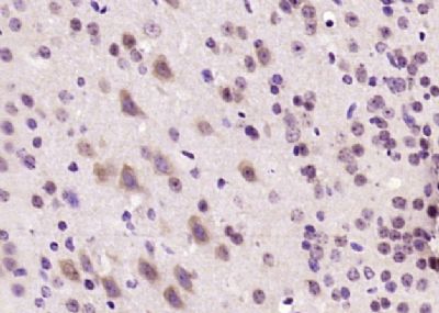
Microcephalin 1/BRIT1 Polyclonal Antibody
Purified Rabbit Polyclonal Antibody (Pab)
- SPECIFICATION
- CITATIONS
- PROTOCOLS
- BACKGROUND

Application
| WB, IHC-P, IHC-F, IF, ICC, E |
|---|---|
| Primary Accession | Q8NEM0 |
| Reactivity | Dog |
| Host | Rabbit |
| Clonality | Polyclonal |
| Calculated MW | 93 KDa |
| Physical State | Liquid |
| Immunogen | KLH conjugated synthetic peptide derived from human Microcephalin 1/BRIT1 |
| Epitope Specificity | 11-110/835 |
| Isotype | IgG |
| Purity | affinity purified by Protein A |
| Buffer | 0.01M TBS (pH7.4) with 1% BSA, 0.02% Proclin300 and 50% Glycerol. |
| SUBCELLULAR LOCATION | Cytoplasm, cytoskeleton, centrosome. |
| SIMILARITY | Contains 3 BRCT domains. |
| SUBUNIT | Contains 3 BRCT domains. |
| DISEASE | Defects in MCPH1 are the cause of microcephaly primary type 1 (MCPH1) [MIM:251200]; also known as true microcephaly or microcephaly vera. Microcephaly is defined as a head circumference more than 3 standard deviations below the age-related mean. Brain weight is markedly reduced and the cerebral cortex is disproportionately small. Despite this marked reduction in size, the gyral pattern is relatively well preserved, with no major abnormality in cortical architecture. Primary microcephaly is further defined by the absence of other syndromic features or significant neurological deficits. This entity is inherited as autosomal recessive trait. |
| Important Note | This product as supplied is intended for research use only, not for use in human, therapeutic or diagnostic applications. |
| Background Descriptions | Microcephalin modulates brain size and has been proliferating under strong positive selection for several thousand years, although the nature of the positive selection is poorly understood. Human Microcephalin contains three BRCA1 C-terminal (BRCT) domains and shares 57% identity with its mouse ortholog, the most conserved regions being BRCT domains where there is 80% identity. Predominant expression of human Microcephalin is observed in fetal brain, liver and kidney tissues and is expressed during neurogenesis in mice. Microcephalin displays significantly higher rates of protein evolution in primates than in rodents; this trend is most noticeable for the subset of genes associated with nervous system development. Microcephalin has a very young, single nucleotide, polymorphism haplotype associated with modern humans; this gene is presumably still evolving in Homo sapiens. It functions in DNA damage response and regulation of cell cycle checkpoints. |
| Gene ID | 79648 |
|---|---|
| Other Names | Microcephalin, MCPH1 (HGNC:6954) |
| Target/Specificity | Expressed in fetal brain, liver and kidney. |
| Dilution | WB=1:500-2000,IHC-P=1:100-500,IHC-F=1:100-500,ICC=1:100-500,IF=1:100-500,ELISA=1:5000-10000 |
| Storage | Store at -20 ℃ for one year. Avoid repeated freeze/thaw cycles. When reconstituted in sterile pH 7.4 0.01M PBS or diluent of antibody the antibody is stable for at least two weeks at 2-4 ℃. |
| Name | MCPH1 (HGNC:6954) |
|---|---|
| Function | Implicated in chromosome condensation and DNA damage induced cellular responses. May play a role in neurogenesis and regulation of the size of the cerebral cortex. |
| Cellular Location | Cytoplasm, cytoskeleton, microtubule organizing center, centrosome |
| Tissue Location | Expressed in fetal brain, liver and kidney. |
Citations (0)

Thousands of laboratories across the world have published research that depended on the performance of antibodies from Abcepta to advance their research. Check out links to articles that cite our products in major peer-reviewed journals, organized by research category.
Submit your citation using an Abcepta antibody to
info@abcepta.com, and receive a free "I Love Antibodies" mug.
info@abcepta.com, and receive a free "I Love Antibodies" mug.

Application Protocols
Provided below are standard protocols that you may find useful for product applications.
Abcepta welcomes feedback from its customers.
If you have used an Abcepta product and would like to share how it has performed, please click on the "Submit Review" button and provide the requested information. Our staff will examine and post your review and contact you if needed.
If you have any additional inquiries please email technical services at tech@abcepta.com.
$ 385.00
Cat# AP54430
Ordering Information
United States
AlbaniaAustraliaAustriaBelgiumBosnia & HerzegovinaBrazilBulgariaCanadaCentral AmericaChinaCroatiaCyprusCzech RepublicDenmarkEstoniaFinlandFranceGermanyGreeceHong KongHungaryIcelandIndiaIndonesiaIrelandIsraelItalyJapanLatviaLithuaniaLuxembourgMacedoniaMalaysiaMaltaMexicoNetherlandsNew ZealandNorwayPakistanPolandPortugalRomaniaSerbiaSingaporeSlovakiaSloveniaSouth AfricaSouth KoreaSpainSwedenSwitzerlandTaiwanTurkeyUnited KingdomUnited StatesVietnamWorldwideOthers
USA Headquarters
(888) 735-7227 / (858) 622-0099 or (858) 875-1900
Other Products
Shipping Information
Domestic orders (in stock items)
Shipped out the same day. Orders placed after 1 PM (PST) will ship out the next business day.
International orders
Contact your local distributors
