


 Foundational characteristics of cancer include proliferation, angiogenesis, migration, evasion of apoptosis, and cellular immortality. Find key markers for these cellular processes and antibodies to detect them.
 The SUMOplot™ Analysis Program predicts and scores sumoylation sites in your protein. SUMOylation is a post-translational modification involved in various cellular processes, such as nuclear-cytosolic transport, transcriptional regulation, apoptosis, protein stability, response to stress, and progression through the cell cycle.
 The Autophagy Receptor Motif Plotter predicts and scores autophagy receptor binding sites in your protein. Identifying proteins connected to this pathway is critical to understanding the role of autophagy in physiological as well as pathological processes such as development, differentiation, neurodegenerative diseases, stress, infection, and cancer.
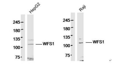
WFS1 Polyclonal Antibody
Purified Rabbit Polyclonal Antibody (Pab)
- SPECIFICATION
- CITATIONS
- PROTOCOLS
- BACKGROUND

Application
| WB, IHC-P, IHC-F, IF, ICC, E |
|---|---|
| Primary Accession | O76024 |
| Reactivity | Rat, Dog, Bovine |
| Host | Rabbit |
| Clonality | Polyclonal |
| Calculated MW | 97 KDa |
| Physical State | Liquid |
| Immunogen | KLH conjugated synthetic peptide derived from human WFS1 |
| Epitope Specificity | 791-890/890 |
| Isotype | IgG |
| Purity | affinity purified by Protein A |
| Buffer | 0.01M TBS (pH7.4) with 1% BSA, 0.02% Proclin300 and 50% Glycerol. |
| SUBCELLULAR LOCATION | Endoplasmic reticulum; endoplasmic reticulum membrane; multipass membrane protein |
| DISEASE | Defects in WFS1 are the cause of Wolfram syndrome type 1 (WFS1) [MIM:222300]. A rare autosomal recessive disorder characterized by juvenile diabetes mellitus, diabetes insipidus, optic atrophy, deafness and various neurological symptoms. Defects in WFS1 are the cause of deafness autosomal dominant type 6 (DFNA6) [MIM:600965]; also called non-syndromic sensorineural deafness autosomal dominant type 14 (DFNA14) or non-syndromic sensorineural deafness autosomal dominant type 38 (DFNA38). DFNA6 is a form of sensorineural hearing loss. Sensorineural deafness results from damage to the neural receptors of the inner ear, the nerve pathways to the brain, or the area of the brain that receives sound information. DFNA6 is a low-frequency hearing loss in which frequencies of 2000 Hz and below are predominantly affected. Many patients have tinnitus, but there are otherwise no associated features such as vertigo. Because high-frequency hearing is generally preserved, patients retain excellent understanding of speech, although presbycusis or noise exposure may cause high-frequency loss later in life. DFNA6 worsens over time without progressing to profound deafness. Defects in WFS1 are the cause of Wolfram-like syndrome autosomal dominant (WFSL) [MIM:614296]. A disease characterized by the clinical triad of congenital progressive hearing impairment, diabetes mellitus, and optic atrophy. The hearing impairment, which is usually diagnosed in the first decade of life, is relatively constant and alters mainly low- and middle-frequency ranges. |
| Important Note | This product as supplied is intended for research use only, not for use in human, therapeutic or diagnostic applications. |
| Background Descriptions | Wolfram syndrome protein (WFS1) is an 890 amino acid protein that contains a cytoplasmic N-terminal domain, followed by nine-transmembrane domains and a luminal C-terminal domain. WFS1 is predominantly localized to the endoplasmic reticulum (ER) (1) and its expression is induced in response to ER stress, partially through transcriptional activation (2,3). Research studies have shown that mutations in the WFS1 gene lead to Wolfram syndrome, an autosomal recessive neurodegenerative disorder defined by young-onset, non-immune, insulin-dependent diabetes mellitus and progressive optic atrophy (4). |
| Gene ID | 7466 |
|---|---|
| Other Names | Wolframin, WFS1 |
| Target/Specificity | Highly expressed in heart followed by brain, placenta, lung and pancreas. Weakly expressed in liver, kidney and skeletal muscle. Also expressed in islet and beta-cell insulinoma cell line. |
| Dilution | WB=1:500-2000,IHC-P=1:100-500,IHC-F=1:100-500,ICC=1:100-500,IF=1:100-500,ELISA=1:5000-10000 |
| Storage | Store at -20 ℃ for one year. Avoid repeated freeze/thaw cycles. When reconstituted in sterile pH 7.4 0.01M PBS or diluent of antibody the antibody is stable for at least two weeks at 2-4 ℃. |
| Name | WFS1 |
|---|---|
| Function | Participates in the regulation of cellular Ca(2+) homeostasis, at least partly, by modulating the filling state of the endoplasmic reticulum Ca(2+) store (PubMed:16989814). Negatively regulates the ER stress response and positively regulates the stability of V-ATPase subunits ATP6V1A and ATP1B1 by preventing their degradation through an unknown proteasome-independent mechanism (PubMed:23035048). |
| Cellular Location | Endoplasmic reticulum membrane; Multi-pass membrane protein. Cytoplasmic vesicle, secretory vesicle. Note=Co-localizes with ATP6V1A in the secretory granules in neuroblastoma cell lines |
| Tissue Location | Highly expressed in heart followed by brain, placenta, lung and pancreas. Weakly expressed in liver, kidney and skeletal muscle. Also expressed in islet and beta-cell insulinoma cell line |
Research Areas
Citations (0)

Thousands of laboratories across the world have published research that depended on the performance of antibodies from Abcepta to advance their research. Check out links to articles that cite our products in major peer-reviewed journals, organized by research category.
Submit your citation using an Abcepta antibody to
info@abcepta.com, and receive a free "I Love Antibodies" mug.
info@abcepta.com, and receive a free "I Love Antibodies" mug.

Application Protocols
Provided below are standard protocols that you may find useful for product applications.
Abcepta welcomes feedback from its customers.
If you have used an Abcepta product and would like to share how it has performed, please click on the "Submit Review" button and provide the requested information. Our staff will examine and post your review and contact you if needed.
If you have any additional inquiries please email technical services at tech@abcepta.com.
$ 385.00
Cat# AP54440
Ordering Information
United States
AlbaniaAustraliaAustriaBelgiumBosnia & HerzegovinaBrazilBulgariaCanadaCentral AmericaChinaCroatiaCyprusCzech RepublicDenmarkEstoniaFinlandFranceGermanyGreeceHong KongHungaryIcelandIndiaIndonesiaIrelandIsraelItalyJapanLatviaLithuaniaLuxembourgMacedoniaMalaysiaMaltaMexicoNetherlandsNew ZealandNorwayPakistanPolandPortugalRomaniaSerbiaSingaporeSlovakiaSloveniaSouth AfricaSouth KoreaSpainSwedenSwitzerlandTaiwanTurkeyUnited KingdomUnited StatesVietnamWorldwideOthers
USA Headquarters
(888) 735-7227 / (858) 622-0099 or (858) 875-1900
Other Products
Shipping Information
Domestic orders (in stock items)
Shipped out the same day. Orders placed after 1 PM (PST) will ship out the next business day.
International orders
Contact your local distributors
