


 Foundational characteristics of cancer include proliferation, angiogenesis, migration, evasion of apoptosis, and cellular immortality. Find key markers for these cellular processes and antibodies to detect them.
 The SUMOplot™ Analysis Program predicts and scores sumoylation sites in your protein. SUMOylation is a post-translational modification involved in various cellular processes, such as nuclear-cytosolic transport, transcriptional regulation, apoptosis, protein stability, response to stress, and progression through the cell cycle.
 The Autophagy Receptor Motif Plotter predicts and scores autophagy receptor binding sites in your protein. Identifying proteins connected to this pathway is critical to understanding the role of autophagy in physiological as well as pathological processes such as development, differentiation, neurodegenerative diseases, stress, infection, and cancer.
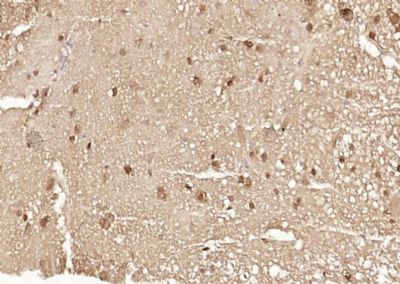
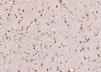
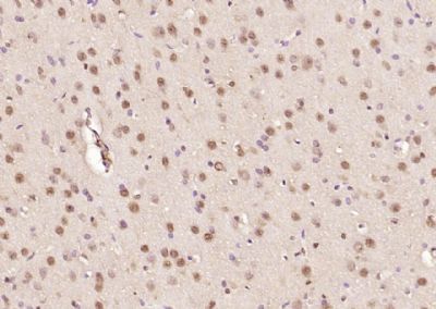
Ataxin 7 Polyclonal Antibody
Purified Rabbit Polyclonal Antibody (Pab)
- SPECIFICATION
- CITATIONS
- PROTOCOLS
- BACKGROUND

Application
| IHC-P, IHC-F, IF, ICC, E |
|---|---|
| Primary Accession | O15265 |
| Reactivity | Rat, Pig, Dog, Bovine |
| Host | Rabbit |
| Clonality | Polyclonal |
| Calculated MW | 95 KDa |
| Physical State | Liquid |
| Immunogen | KLH conjugated synthetic peptide derived from human Ataxin 7 |
| Epitope Specificity | 301-400/892 |
| Isotype | IgG |
| Purity | affinity purified by Protein A |
| Buffer | 0.01M TBS (pH7.4) with 1% BSA, 0.02% Proclin300 and 50% Glycerol. |
| SUBCELLULAR LOCATION | Cytoplasmic (isoform b) and Nuclear (isoform a) |
| SIMILARITY | Belongs to the ataxin-7 family. Contains 1 SCA7 domain. |
| SUBUNIT | Component of the STAGA transcription coactivator-HAT complex, at least composed of SUPT3H, GCN5L2, TAF5L, TAF6L, SUPT7L, TADA3L, TAD1L, TAF10, TAF12, TRRAP, TAF9 and ATXN7. The STAGA core complex is associated with a subcomplex required for histone deubiquitination composed of ATXN7L3, ENY2 and USP22. Interacts with SORBS1, PSMC1 and CRX. Interacts with TRRAP, GCN5L2 and TAF10. Interacts with alpha tubulin. |
| Post-translational modifications | Proteolytically cleaved. The cleavage may be involved in SCA7 degeneration: the isoform fragments may exert distinct toxic influences that could contribute to selective neurodegeneration.Sumoylation decreases the aggregation propensity and cellular toxicity of forms with an expanded poly-Gln region but has no effect on subcellular location or interaction with components of the STAGA complex. |
| DISEASE | Defects in ATXN7 are the cause of spinocerebellar ataxia type 7 (SCA7) [MIM:164500]; also known as olivopontocerebellar atrophy III (OPCA III or OPCA3) or olivopontocerebellar atrophy with retinal degeneration. Spinocerebellar ataxia is a clinically and genetically heterogeneous group of cerebellar disorders. Patients show progressive incoordination of gait and often poor coordination of hands, speech and eye movements, due to degeneration of the cerebellum with variable involvement of the brainstem and spinal cord. SCA7 belongs to the autosomal dominant cerebellar ataxias type II (ADCA II) which are characterized by cerebellar ataxia with retinal degeneration and pigmentary macular dystrophy. |
| Important Note | This product as supplied is intended for research use only, not for use in human, therapeutic or diagnostic applications. |
| Background Descriptions | The human ataxin-7 gene, also known as spinocerebellar ataxia 7 or SCA7, maps to chromosome 3p13-p12, has a 2,727-bp open reading frame, and encodes a 892 amino acid protein containing a nuclear localization signal and a polyglutamine tract (1,2). SCA7 is an autosomal dominant neurodegenerative disorder characterized by ataxia and selective neuronal cell loss caused by the expansion of a translated CAG repeat encoding a polyglutamine tract in ataxin-7, which is the SCA7 gene product (3,4). Ataxin-7 is expressed within neurons both affected and unaffected in SCA7 pathology with subcellular localization being variable depending upon the neuronal subtype (5). Polyglutamine expanded in ataxin-7 may carry out its pathogenic effects in the nucleus by altering the matrix-associated nuclear structure and/or by disrupting nucleolar function (6). |
| Gene ID | 6314 |
|---|---|
| Other Names | Ataxin-7, Spinocerebellar ataxia type 7 protein, ATXN7, SCA7 |
| Target/Specificity | Isoform a and isoform b are expressed in CNS, but isoform a is expressed predominantly in the peripherical tissues. Isoform b is also highly expressed in the frontal lobe, skeletal muscle and spinal cord and is expressed at a lower level in the lung, lymphoblast and intestine. |
| Dilution | IHC-P=1:100-500,IHC-F=1:100-500,ICC=1:100-500,IF=1:100-500,ELISA=1:5000-10000 |
| Storage | Store at -20 ℃ for one year. Avoid repeated freeze/thaw cycles. When reconstituted in sterile pH 7.4 0.01M PBS or diluent of antibody the antibody is stable for at least two weeks at 2-4 ℃. |
| Name | ATXN7 |
|---|---|
| Synonyms | SCA7 {ECO:0000303|PubMed:9288099} |
| Function | Acts as a component of the SAGA (aka STAGA) transcription coactivator-HAT complex (PubMed:15932940, PubMed:18206972). Mediates the interaction of SAGA complex with the CRX and is involved in CRX- dependent gene activation (PubMed:15932940, PubMed:18206972). Probably involved in tethering the deubiquitination module within the SAGA complex (PubMed:24493646). Necessary for microtubule cytoskeleton stabilization (PubMed:22100762). Involved in neurodegeneration (PubMed:9288099). |
| Cellular Location | [Isoform a]: Nucleus. Nucleus, nucleolus. Nucleus matrix. Cytoplasm, cytoskeleton. Note=In addition to a diffuse distribution throughout the nucleus, it is associated with the nuclear matrix and the nucleolus (PubMed:10441328). It is able to shuttle between the nucleus and cytoplasm (PubMed:16314424) |
| Tissue Location | [Isoform a]: Isoform a is expressed in CNS, but is expressed predominantly in the peripherical tissues |
Research Areas
Citations (0)

Thousands of laboratories across the world have published research that depended on the performance of antibodies from Abcepta to advance their research. Check out links to articles that cite our products in major peer-reviewed journals, organized by research category.
Submit your citation using an Abcepta antibody to
info@abcepta.com, and receive a free "I Love Antibodies" mug.
info@abcepta.com, and receive a free "I Love Antibodies" mug.

Application Protocols
Provided below are standard protocols that you may find useful for product applications.
Abcepta welcomes feedback from its customers.
If you have used an Abcepta product and would like to share how it has performed, please click on the "Submit Review" button and provide the requested information. Our staff will examine and post your review and contact you if needed.
If you have any additional inquiries please email technical services at tech@abcepta.com.
$ 385.00
Cat# AP54452
Ordering Information
United States
AlbaniaAustraliaAustriaBelgiumBosnia & HerzegovinaBrazilBulgariaCanadaCentral AmericaChinaCroatiaCyprusCzech RepublicDenmarkEstoniaFinlandFranceGermanyGreeceHong KongHungaryIcelandIndiaIndonesiaIrelandIsraelItalyJapanLatviaLithuaniaLuxembourgMacedoniaMalaysiaMaltaMexicoNetherlandsNew ZealandNorwayPakistanPolandPortugalRomaniaSerbiaSingaporeSlovakiaSloveniaSouth AfricaSouth KoreaSpainSwedenSwitzerlandTaiwanTurkeyUnited KingdomUnited StatesVietnamWorldwideOthers
USA Headquarters
(888) 735-7227 / (858) 622-0099 or (858) 875-1900
Shipping Information
Domestic orders (in stock items)
Shipped out the same day. Orders placed after 1 PM (PST) will ship out the next business day.
International orders
Contact your local distributors
