


 Foundational characteristics of cancer include proliferation, angiogenesis, migration, evasion of apoptosis, and cellular immortality. Find key markers for these cellular processes and antibodies to detect them.
 The SUMOplot™ Analysis Program predicts and scores sumoylation sites in your protein. SUMOylation is a post-translational modification involved in various cellular processes, such as nuclear-cytosolic transport, transcriptional regulation, apoptosis, protein stability, response to stress, and progression through the cell cycle.
 The Autophagy Receptor Motif Plotter predicts and scores autophagy receptor binding sites in your protein. Identifying proteins connected to this pathway is critical to understanding the role of autophagy in physiological as well as pathological processes such as development, differentiation, neurodegenerative diseases, stress, infection, and cancer.
> home > Products > Primary Antibodies > Antibody Collections > Catalog Updated > ATP11C Antibody (Center)
ATP11C Antibody (Center)
Affinity Purified Rabbit Polyclonal Antibody (Pab)
- SPECIFICATION
- CITATIONS: 1
- PROTOCOLS
- BACKGROUND

Application
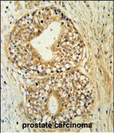
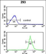
| FC, IHC-P, WB, E |
|---|---|
| Primary Accession | Q8NB49 |
| Other Accession | NP_001010986.1 |
| Reactivity | Human, Hamster, Mouse |
| Host | Rabbit |
| Clonality | Polyclonal |
| Isotype | Rabbit IgG |
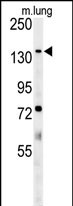
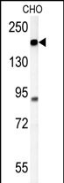
| Calculated MW | 129477 Da |
| Antigen Region | 589-616 aa |
| Gene ID | 286410 |
|---|---|
| Other Names | Phospholipid-transporting ATPase IG, ATPase IQ, ATPase class VI type 11C, P4-ATPase flippase complex alpha subunit ATP11C, ATP11C, ATPIG, ATPIQ |
| Target/Specificity | This ATP11C antibody is generated from rabbits immunized with a KLH conjugated synthetic peptide between 589-616 amino acids from the Central region of human ATP11C. |
| Dilution | FC~~1:10~50 IHC-P~~1:50~100 WB~~1:1000 E~~Use at an assay dependent concentration. |
| Format | Purified polyclonal antibody supplied in PBS with 0.09% (W/V) sodium azide. This antibody is purified through a protein A column, followed by peptide affinity purification. |
| Storage | Maintain refrigerated at 2-8°C for up to 2 weeks. For long term storage store at -20°C in small aliquots to prevent freeze-thaw cycles. |
| Precautions | ATP11C Antibody (Center) is for research use only and not for use in diagnostic or therapeutic procedures. |
| Name | ATP11C {ECO:0000303|PubMed:26944472} |
|---|---|
| Synonyms | ATPIG, ATPIQ |
| Function | Catalytic component of a P4-ATPase flippase complex which catalyzes the hydrolysis of ATP coupled to the transport of aminophospholipids, phosphatidylserines (PS) and phosphatidylethanolamines (PE), from the outer to the inner leaflet of the plasma membrane (PubMed:24904167, PubMed:25315773, PubMed:26567335, PubMed:32493773). Major PS-flippase in immune cell subsets. In erythrocyte plasma membrane, it is required to maintain PS in the inner leaflet preventing its exposure on the surface. This asymmetric distribution is critical for the survival of erythrocytes in circulation since externalized PS is a phagocytic signal for erythrocyte clearance by splenic macrophages (PubMed:26944472). Required for B cell differentiation past the pro-B cell stage (By similarity). Seems to mediate PS flipping in pro-B cells (By similarity). May be involved in the transport of cholestatic bile acids (By similarity). |
| Cellular Location | Cell membrane; Multi-pass membrane protein. Endoplasmic reticulum membrane; Multi-pass membrane protein. Early endosome membrane; Multi-pass membrane protein. Recycling endosome membrane; Multi-pass membrane protein. Note=Efficient exit from the endoplasmic reticulum requires the presence of TMEM30A. Internalized via clathrin-dependent endocytosis in response to ca(2+) signaling induced by G-protein coupled serotonin and histamine receptors |
| Tissue Location | Widely expressed. |
Research Areas
Citations ( 0 )

Application Protocols
Provided below are standard protocols that you may find useful for product applications.
Background
The function of ATP11C remains unknown.
References
Matsuoka, S., et al. Science 316(5828):1160-1166(2007)
Andrew Nesbit, M., et al. Genomics 84(6):1060-1070(2004)
Halleck, M.S., et al. Physiol. Genomics 1(3):139-150(1999)
Yokoi, H., et al. Genomics 20(3):404-411(1994)
Abcepta welcomes feedback from its customers.
If you have used an Abcepta product and would like to share how it has performed, please click on the "Submit Review" button and provide the requested information. Our staff will examine and post your review and contact you if needed.
If you have any additional inquiries please email technical services at tech@abcepta.com.
$ 150.00
$ 385.00
Cat# AP5446c
Ordering Information
United States
AlbaniaAustraliaAustriaBelgiumBosnia & HerzegovinaBrazilBulgariaCanadaCentral AmericaChinaCroatiaCyprusCzech RepublicDenmarkEstoniaFinlandFranceGermanyGreeceHong KongHungaryIcelandIndiaIndonesiaIrelandIsraelItalyJapanLatviaLithuaniaLuxembourgMacedoniaMalaysiaMaltaMexicoNetherlandsNew ZealandNorwayPakistanPolandPortugalRomaniaSerbiaSingaporeSlovakiaSloveniaSouth AfricaSouth KoreaSpainSwedenSwitzerlandTaiwanTurkeyUnited KingdomUnited StatesVietnamWorldwideOthers
USA Headquarters
(888) 735-7227 / (858) 622-0099 or (858) 875-1900
Other Products
Shipping Information
Domestic orders (in stock items)
Shipped out the same day. Orders placed after 1 PM (PST) will ship out the next business day.
International orders
Contact your local distributors
