


 Foundational characteristics of cancer include proliferation, angiogenesis, migration, evasion of apoptosis, and cellular immortality. Find key markers for these cellular processes and antibodies to detect them.
 The SUMOplot™ Analysis Program predicts and scores sumoylation sites in your protein. SUMOylation is a post-translational modification involved in various cellular processes, such as nuclear-cytosolic transport, transcriptional regulation, apoptosis, protein stability, response to stress, and progression through the cell cycle.
 The Autophagy Receptor Motif Plotter predicts and scores autophagy receptor binding sites in your protein. Identifying proteins connected to this pathway is critical to understanding the role of autophagy in physiological as well as pathological processes such as development, differentiation, neurodegenerative diseases, stress, infection, and cancer.
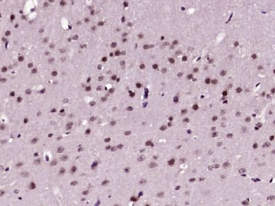
HES4 Polyclonal Antibody
Purified Rabbit Polyclonal Antibody (Pab)
- SPECIFICATION
- CITATIONS
- PROTOCOLS
- BACKGROUND

Application
| WB, IHC-P, IHC-F, IF, ICC, E |
|---|---|
| Primary Accession | Q9HCC6 |
| Reactivity | Rat, Pig, Dog |
| Host | Rabbit |
| Clonality | Polyclonal |
| Calculated MW | 24 KDa |
| Physical State | Liquid |
| Immunogen | KLH conjugated synthetic peptide derived from human HES4 |
| Epitope Specificity | 1-100/221 |
| Isotype | IgG |
| Purity | affinity purified by Protein A |
| Buffer | 0.01M TBS (pH7.4) with 1% BSA, 0.02% Proclin300 and 50% Glycerol. |
| SUBCELLULAR LOCATION | Nuclear |
| SIMILARITY | Contains 1 bHLH (basic helix-loop-helix) domain. Contains 1 Orange domain. |
| SUBUNIT | Transcription repression requires formation of a complex with a corepressor protein of the Groucho/TLE family |
| Important Note | This product as supplied is intended for research use only, not for use in human, therapeutic or diagnostic applications. |
| Background Descriptions | The Drosophila hairy and Enhancer of split genes encode basic helix-loop-helix (bHLH) transcriptional repressors that function in the Notch signaling pathway and control segmentation and neural development during embryogenesis. The mammalian homologues of Drosophila hairy and Enhancer of split are the HES gene family members, HES1-6, which also encode bHLH transcriptional repressors that regulate myogenesis and neurogenesis. The HES family members form a complex with TLE, the mammalian homologue of Groucho, and this interaction is mediated by the carboxy terminal WRPW motif of the HES proteins. The HES/TLE complex functions by directly binding to DNA, instead of interfering with activator proteins. Most HES family members, including HES1 and HES5, preferentially bind to the N box (CACNAG) as opposed to the E box (CANNTG). HES2 binds to both N and E box sites, while HES6 does not bind DNA. Rather, HES6 inhibits HES1 activity, thereby promoting transcription. HES1 and HES2 are expressed in a variety of adult and embryonic tissues. HES3 is expressed exclusively in cerebellar Purkinje cells, and HES5 is found solely in the nervous system. HES6 is produced in brain as well as in the limb buds of developing embryos. |
| Gene ID | 57801 |
|---|---|
| Other Names | Transcription factor HES-4, hHES4, Class B basic helix-loop-helix protein 42, bHLHb42, Hairy and enhancer of split 4, bHLH factor Hes4, HES4, BHLHB42 |
| Dilution | WB=1:500-2000,IHC-P=1:100-500,IHC-F=1:100-500,ICC=1:100-500,IF=1:100-500,ELISA=1:5000-10000 |
| Storage | Store at -20 ℃ for one year. Avoid repeated freeze/thaw cycles. When reconstituted in sterile pH 7.4 0.01M PBS or diluent of antibody the antibody is stable for at least two weeks at 2-4 ℃. |
| Name | HES4 |
|---|---|
| Synonyms | BHLHB42 |
| Function | Transcriptional repressor. Binds DNA on N-box motifs: 5'- CACNAG-3' (By similarity). |
| Cellular Location | Nucleus {ECO:0000255|PROSITE-ProRule:PRU00380, ECO:0000255|PROSITE-ProRule:PRU00981} |
Research Areas
Citations (0)

Thousands of laboratories across the world have published research that depended on the performance of antibodies from Abcepta to advance their research. Check out links to articles that cite our products in major peer-reviewed journals, organized by research category.
Submit your citation using an Abcepta antibody to
info@abcepta.com, and receive a free "I Love Antibodies" mug.
info@abcepta.com, and receive a free "I Love Antibodies" mug.

Application Protocols
Provided below are standard protocols that you may find useful for product applications.
Abcepta welcomes feedback from its customers.
If you have used an Abcepta product and would like to share how it has performed, please click on the "Submit Review" button and provide the requested information. Our staff will examine and post your review and contact you if needed.
If you have any additional inquiries please email technical services at tech@abcepta.com.
$ 385.00
Cat# AP54549
Ordering Information
United States
AlbaniaAustraliaAustriaBelgiumBosnia & HerzegovinaBrazilBulgariaCanadaCentral AmericaChinaCroatiaCyprusCzech RepublicDenmarkEstoniaFinlandFranceGermanyGreeceHong KongHungaryIcelandIndiaIndonesiaIrelandIsraelItalyJapanLatviaLithuaniaLuxembourgMacedoniaMalaysiaMaltaMexicoNetherlandsNew ZealandNorwayPakistanPolandPortugalRomaniaSerbiaSingaporeSlovakiaSloveniaSouth AfricaSouth KoreaSpainSwedenSwitzerlandTaiwanTurkeyUnited KingdomUnited StatesVietnamWorldwideOthers
USA Headquarters
(888) 735-7227 / (858) 622-0099 or (858) 875-1900
Shipping Information
Domestic orders (in stock items)
Shipped out the same day. Orders placed after 1 PM (PST) will ship out the next business day.
International orders
Contact your local distributors
