


 Foundational characteristics of cancer include proliferation, angiogenesis, migration, evasion of apoptosis, and cellular immortality. Find key markers for these cellular processes and antibodies to detect them.
 The SUMOplot™ Analysis Program predicts and scores sumoylation sites in your protein. SUMOylation is a post-translational modification involved in various cellular processes, such as nuclear-cytosolic transport, transcriptional regulation, apoptosis, protein stability, response to stress, and progression through the cell cycle.
 The Autophagy Receptor Motif Plotter predicts and scores autophagy receptor binding sites in your protein. Identifying proteins connected to this pathway is critical to understanding the role of autophagy in physiological as well as pathological processes such as development, differentiation, neurodegenerative diseases, stress, infection, and cancer.
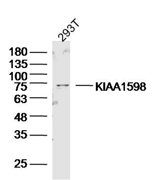
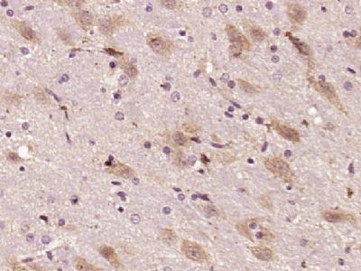
KIAA1598 Polyclonal Antibody
Purified Rabbit Polyclonal Antibody (Pab)
- SPECIFICATION
- CITATIONS
- PROTOCOLS
- BACKGROUND

Application
| IHC-P, IHC-F, IF, ICC |
|---|---|
| Primary Accession | A0MZ66 |
| Reactivity | Rat, Pig, Bovine |
| Host | Rabbit |
| Clonality | Polyclonal |
| Calculated MW | 71640 Da |
| Gene ID | 57698 |
|---|---|
| Other Names | Shootin-1 {ECO:0000312|HGNC:HGNC:29319}, Shootin1, SHTN1 (HGNC:29319), KIAA1598 |
| Dilution | Elisa=1:500-1000,IHC-P=1:100-500,IHC-F=1:100-500,IF=1:100-500,ICC=1:100-500, |
| Format | 0.01M TBS(pH7.4), 0.09% (W/V) sodium azide and 50% Glyce |
| Storage | Store at -20 ℃ for one year. Avoid repeated freeze/thaw cycles. When reconstituted in sterile pH 7.4 0.01M PBS or diluent of antibody the antibody is stable for at least two weeks at 2-4 ℃. |
| Name | SHTN1 (HGNC:29319) |
|---|---|
| Synonyms | KIAA1598 |
| Function | Involved in the generation of internal asymmetric signals required for neuronal polarization and neurite outgrowth. Mediates netrin-1-induced F-actin-substrate coupling or 'clutch engagement' within the axon growth cone through activation of CDC42, RAC1 and PAK1- dependent signaling pathway, thereby converting the F-actin retrograde flow into traction forces, concomitantly with filopodium extension and axon outgrowth. Plays a role in cytoskeletal organization by regulating the subcellular localization of phosphoinositide 3-kinase (PI3K) activity at the axonal growth cone. Also plays a role in regenerative neurite outgrowth. In the developing cortex, cooperates with KIF20B to promote both the transition from the multipolar to the bipolar stage and the radial migration of cortical neurons from the ventricular zone toward the superficial layer of the neocortex. Involved in the accumulation of phosphatidylinositol 3,4,5-trisphosphate (PIP3) in the growth cone of primary hippocampal neurons. |
| Cellular Location | Perikaryon {ECO:0000250|UniProtKB:Q8K2Q9}. Cell projection, axon {ECO:0000250|UniProtKB:Q8K2Q9}. Cell projection, growth cone {ECO:0000250|UniProtKB:Q8K2Q9}. Cytoplasm, cytoskeleton {ECO:0000250|UniProtKB:Q8K2Q9}. Cell projection, filopodium {ECO:0000250|UniProtKB:A0MZ67}. Cell projection, lamellipodium {ECO:0000250|UniProtKB:A0MZ67}. Note=Localizes in multiple growth cones at neurite tips before the neuronal symmetry-breaking step. Accumulates in growth cones of a single nascent axon in a neurite length-dependent manner during the neuronal symmetry-breaking step; when absent from the nascent axon's siblings, probably due to competitive transport, prevents the formation of surplus axons. Transported anterogradely from the soma to the axon growth cone in an actin and myosin-dependent manner and passively diffuses back to the cell bodies. Colocalized with L1CAM in close apposition with actin filaments in filopodia and lamellipodia of axonal growth cones in hippocampal neurons. Exhibits retrograde movements in filopodia and lamellopodia of axonal growth cones. Colocalized with KIF20B along microtubules to the tip of the growing cone in primary hippocampal neurons. Recruited to the growth cone of developing axon in a KIF20B- and microtubule-dependent manner {ECO:0000250|UniProtKB:A0MZ67, ECO:0000250|UniProtKB:Q8K2Q9} |
Research Areas
Citations (0)

Thousands of laboratories across the world have published research that depended on the performance of antibodies from Abcepta to advance their research. Check out links to articles that cite our products in major peer-reviewed journals, organized by research category.
Submit your citation using an Abcepta antibody to
info@abcepta.com, and receive a free "I Love Antibodies" mug.
info@abcepta.com, and receive a free "I Love Antibodies" mug.

Application Protocols
Provided below are standard protocols that you may find useful for product applications.
Abcepta welcomes feedback from its customers.
If you have used an Abcepta product and would like to share how it has performed, please click on the "Submit Review" button and provide the requested information. Our staff will examine and post your review and contact you if needed.
If you have any additional inquiries please email technical services at tech@abcepta.com.
$ 350.00
Cat# AP54652
Ordering Information
United States
AlbaniaAustraliaAustriaBelgiumBosnia & HerzegovinaBrazilBulgariaCanadaCentral AmericaChinaCroatiaCyprusCzech RepublicDenmarkEstoniaFinlandFranceGermanyGreeceHong KongHungaryIcelandIndiaIndonesiaIrelandIsraelItalyJapanLatviaLithuaniaLuxembourgMacedoniaMalaysiaMaltaNetherlandsNew ZealandNorwayPakistanPolandPortugalRomaniaSerbiaSingaporeSlovakiaSloveniaSouth AfricaSouth KoreaSpainSwedenSwitzerlandTaiwanTurkeyUnited KingdomUnited StatesVietnamWorldwideOthers
USA Headquarters
(888) 735-7227 / (858) 622-0099 or (858) 875-1900
Other Products
Shipping Information
Domestic orders (in stock items)
Shipped out the same day. Orders placed after 1 PM (PST) will ship out the next business day.
International orders
Contact your local distributors
