


 Foundational characteristics of cancer include proliferation, angiogenesis, migration, evasion of apoptosis, and cellular immortality. Find key markers for these cellular processes and antibodies to detect them.
 The SUMOplot™ Analysis Program predicts and scores sumoylation sites in your protein. SUMOylation is a post-translational modification involved in various cellular processes, such as nuclear-cytosolic transport, transcriptional regulation, apoptosis, protein stability, response to stress, and progression through the cell cycle.
 The Autophagy Receptor Motif Plotter predicts and scores autophagy receptor binding sites in your protein. Identifying proteins connected to this pathway is critical to understanding the role of autophagy in physiological as well as pathological processes such as development, differentiation, neurodegenerative diseases, stress, infection, and cancer.
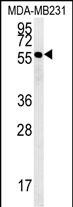
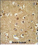
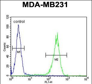
RAB11FIP2 Antibody (Center)
Affinity Purified Rabbit Polyclonal Antibody (Pab)
- SPECIFICATION
- CITATIONS
- PROTOCOLS
- BACKGROUND

Application
| FC, IHC-P, WB, E |
|---|---|
| Primary Accession | Q7L804 |
| Other Accession | NP_055719.1 |
| Reactivity | Human |
| Host | Rabbit |
| Clonality | Polyclonal |
| Isotype | Rabbit IgG |
| Calculated MW | 58279 Da |
| Antigen Region | 345-374 aa |
| Gene ID | 22841 |
|---|---|
| Other Names | Rab11 family-interacting protein 2, Rab11-FIP2, NRip11, RAB11FIP2, KIAA0941 |
| Target/Specificity | This RAB11FIP2 antibody is generated from rabbits immunized with a KLH conjugated synthetic peptide between 345-374 amino acids from the Central region of human RAB11FIP2. |
| Dilution | FC~~1:10~50 IHC-P~~1:50~100 WB~~1:1000 E~~Use at an assay dependent concentration. |
| Format | Purified polyclonal antibody supplied in PBS with 0.09% (W/V) sodium azide. This antibody is purified through a protein A column, followed by peptide affinity purification. |
| Storage | Maintain refrigerated at 2-8°C for up to 2 weeks. For long term storage store at -20°C in small aliquots to prevent freeze-thaw cycles. |
| Precautions | RAB11FIP2 Antibody (Center) is for research use only and not for use in diagnostic or therapeutic procedures. |
| Name | RAB11FIP2 |
|---|---|
| Synonyms | KIAA0941 |
| Function | A Rab11 effector binding preferentially phosphatidylinositol 3,4,5-trisphosphate (PtdInsP3) and phosphatidic acid (PA) and acting in the regulation of the transport of vesicles from the endosomal recycling compartment (ERC) to the plasma membrane. Involved in insulin granule exocytosis. Also involved in receptor-mediated endocytosis and membrane trafficking of recycling endosomes, probably originating from clathrin-coated vesicles. Required in a complex with MYO5B and RAB11 for the transport of NPC1L1 to the plasma membrane. Also acts as a regulator of cell polarity. Plays an essential role in phagocytosis through a mechanism involving TICAM2, RAC1 and CDC42 Rho GTPases for controlling actin-dynamics. |
| Cellular Location | Cell projection, phagocytic cup. Cell membrane; Peripheral membrane protein. Recycling endosome membrane; Peripheral membrane protein Note=Translocates with RAB11A from the vesicles of the endocytic recycling compartment (ERC) to the plasma membrane |

Thousands of laboratories across the world have published research that depended on the performance of antibodies from Abcepta to advance their research. Check out links to articles that cite our products in major peer-reviewed journals, organized by research category.
info@abcepta.com, and receive a free "I Love Antibodies" mug.

Provided below are standard protocols that you may find useful for product applications.
Background
RAB11FIP2 is an adapter protein that plays a role in the secretory pathway. It is thought to be important for endosome recycling and receptor-mediated endocytosis. In endosome recycling, RAB11-FIP2 regulates vesicle transport from the endosomal recycling compartment (ERC) to the plasma membrane.
References
Wang, Z., et al. Cell 135(3):535-548(2008)
Utley, T.J., et al. Proc. Natl. Acad. Sci. U.S.A. 105(29):10209-10214(2008)
Ducharme, N.A., et al. Am. J. Physiol., Cell Physiol. 293 (3), C1059-C1072 (2007)
If you have used an Abcepta product and would like to share how it has performed, please click on the "Submit Review" button and provide the requested information. Our staff will examine and post your review and contact you if needed.
If you have any additional inquiries please email technical services at tech@abcepta.com.
Ordering Information
Shipping Information
