


 Foundational characteristics of cancer include proliferation, angiogenesis, migration, evasion of apoptosis, and cellular immortality. Find key markers for these cellular processes and antibodies to detect them.
 The SUMOplot™ Analysis Program predicts and scores sumoylation sites in your protein. SUMOylation is a post-translational modification involved in various cellular processes, such as nuclear-cytosolic transport, transcriptional regulation, apoptosis, protein stability, response to stress, and progression through the cell cycle.
 The Autophagy Receptor Motif Plotter predicts and scores autophagy receptor binding sites in your protein. Identifying proteins connected to this pathway is critical to understanding the role of autophagy in physiological as well as pathological processes such as development, differentiation, neurodegenerative diseases, stress, infection, and cancer.
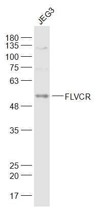
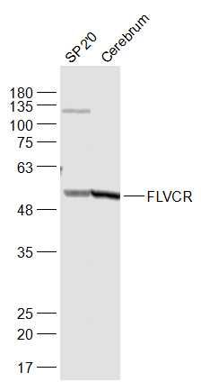
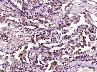
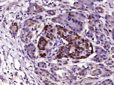
FLVCR Polyclonal Antibody
Purified Rabbit Polyclonal Antibody (Pab)
- SPECIFICATION
- CITATIONS
- PROTOCOLS
- BACKGROUND

Application
| WB, IHC-P, IHC-F, IF, ICC, E |
|---|---|
| Primary Accession | Q9Y5Y0 |
| Reactivity | Rat, Pig |
| Host | Rabbit |
| Clonality | Polyclonal |
| Calculated MW | 60 KDa |
| Physical State | Liquid |
| Immunogen | KLH conjugated synthetic peptide derived from Human FLVCR |
| Epitope Specificity | 451-550/555 |
| Isotype | IgG |
| Purity | affinity purified by Protein A |
| Buffer | 0.01M TBS (pH7.4) with 1% BSA, 0.02% Proclin300 and 50% Glycerol. |
| SUBCELLULAR LOCATION | Cell membrane. |
| SIMILARITY | Belongs to the major facilitator superfamily. Feline leukemia virus subgroup C receptor (TC 2.A.1.28.1) family. |
| SUBUNIT | Interacts with HPX. |
| DISEASE | Defects in FLVCR1 are the cause of posterior column ataxia with retinitis pigmentosa (PCARP) [MIM:609033]. A neurodegenerative syndrome beginning in infancy with areflexia and retinitis pigmentosa. Nyctalopia (night blindness) and peripheral visual field loss are usually evident during late childhood or teenage years, with subsequent progressive constriction of the visual fields and loss of central retinal function over time. A sensory ataxia caused by degeneration of the posterior columns of the spinal cord results in a loss of proprioceptive sensation that is clinically evident in the second decade of life and gradually progresses. Scoliosis, camptodactyly, achalasia, gastrointestinal dysmotility, and a sensory peripheral neuropathy are variable features of the disease. Affected individuals have no clinical or radiological evidence of cerebral or cerebellar involvement. Note=Defective neuronal heme transmembrane export due to FLVCR1 mutations may abrogate the neuroprotective effects of neuroglobin and initiate an apoptotic cascade that results in the selective degeneration of photoreceptors in the neurosensory retina and sensory neurons in the posterior spinal cord. |
| Important Note | This product as supplied is intended for research use only, not for use in human, therapeutic or diagnostic applications. |
| Background Descriptions | FLVCR is responsible for the exportation of cytoplasmic heme groups. It is believed that it may protect developing erythroid cells from heme toxicity. Expression of FLVCR in cells will cause susceptibility to FeLV-C (Feline leukemia virus subgroup C) in vitro. FLVCR is found in all hematopoietic tissues, including peripheral blood lymphocytes and fetal liver, and some expression is found in pancreas and kidney. It is down-regulated in haemopoietic progenitor cells undergoing differentiation and hemoglobinization. |
| Gene ID | 28982 |
|---|---|
| Other Names | Feline leukemia virus subgroup C receptor-related protein 1, Feline leukemia virus subgroup C receptor, hFLVCR, FLVCR1, FLVCR |
| Target/Specificity | Found all hematopoietic tissues including peripheral blood lymphocytes. Some expression is found in pancreas and kidney. |
| Dilution | WB=1:500-2000,IHC-P=1:100-500,IHC-F=1:100-500,ICC=1:100-500,IF=1:100-500,ELISA=1:5000-10000 |
| Storage | Store at -20 ℃ for one year. Avoid repeated freeze/thaw cycles. When reconstituted in sterile pH 7.4 0.01M PBS or diluent of antibody the antibody is stable for at least two weeks at 2-4 ℃. |
| Name | FLVCR1 {ECO:0000303|PubMed:16439531, ECO:0000312|HGNC:HGNC:24682} |
|---|---|
| Function | Uniporter that mediates the transport of extracellular choline and ethanolamine into cells, thereby playing a key role in phospholipid biosynthesis (PubMed:37100056, PubMed:38693265, PubMed:38778100, PubMed:39306721). Choline and ethanolamine are the precursors of phosphatidylcholine and phosphatidylethanolamine, respectively, the two most abundant phospholipids (PubMed:38693265, PubMed:38778100). Transport is not coupled with proton transport and is exclusively driven by the choline (or ethanolamine) gradient across the plasma membrane (PubMed:38693265, PubMed:38778100). Also acts as a heme b transporter that mediates heme efflux from the cytoplasm to the extracellular compartment (PubMed:15369674, PubMed:20610401, PubMed:22483575, PubMed:23187127, PubMed:27923065). |
| Cellular Location | [Isoform 1]: Cell membrane; Multi-pass membrane protein |
| Tissue Location | Found all hematopoietic tissues including peripheral blood lymphocytes. Some expression is found in pancreas and kidney. |
Research Areas
Citations (0)

Thousands of laboratories across the world have published research that depended on the performance of antibodies from Abcepta to advance their research. Check out links to articles that cite our products in major peer-reviewed journals, organized by research category.
Submit your citation using an Abcepta antibody to
info@abcepta.com, and receive a free "I Love Antibodies" mug.
info@abcepta.com, and receive a free "I Love Antibodies" mug.

Application Protocols
Provided below are standard protocols that you may find useful for product applications.
Abcepta welcomes feedback from its customers.
If you have used an Abcepta product and would like to share how it has performed, please click on the "Submit Review" button and provide the requested information. Our staff will examine and post your review and contact you if needed.
If you have any additional inquiries please email technical services at tech@abcepta.com.
$ 385.00
Cat# AP54831
Ordering Information
United States
AlbaniaAustraliaAustriaBelgiumBosnia & HerzegovinaBrazilBulgariaCanadaCentral AmericaChinaCroatiaCyprusCzech RepublicDenmarkEstoniaFinlandFranceGermanyGreeceHong KongHungaryIcelandIndiaIndonesiaIrelandIsraelItalyJapanLatviaLithuaniaLuxembourgMacedoniaMalaysiaMaltaMexicoNetherlandsNew ZealandNorwayPakistanPolandPortugalRomaniaSerbiaSingaporeSlovakiaSloveniaSouth AfricaSouth KoreaSpainSwedenSwitzerlandTaiwanTurkeyUnited KingdomUnited StatesVietnamWorldwideOthers
USA Headquarters
(888) 735-7227 / (858) 622-0099 or (858) 875-1900
Other Products
Shipping Information
Domestic orders (in stock items)
Shipped out the same day. Orders placed after 1 PM (PST) will ship out the next business day.
International orders
Contact your local distributors
