


 Foundational characteristics of cancer include proliferation, angiogenesis, migration, evasion of apoptosis, and cellular immortality. Find key markers for these cellular processes and antibodies to detect them.
 The SUMOplot™ Analysis Program predicts and scores sumoylation sites in your protein. SUMOylation is a post-translational modification involved in various cellular processes, such as nuclear-cytosolic transport, transcriptional regulation, apoptosis, protein stability, response to stress, and progression through the cell cycle.
 The Autophagy Receptor Motif Plotter predicts and scores autophagy receptor binding sites in your protein. Identifying proteins connected to this pathway is critical to understanding the role of autophagy in physiological as well as pathological processes such as development, differentiation, neurodegenerative diseases, stress, infection, and cancer.
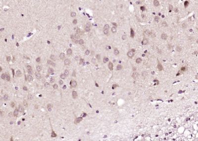
Prickle Polyclonal Antibody
Purified Rabbit Polyclonal Antibody (Pab)
- SPECIFICATION
- CITATIONS
- PROTOCOLS
- BACKGROUND

Application
| IHC-P, IHC-F, IF, ICC, E |
|---|---|
| Primary Accession | Q96MT3 |
| Reactivity | Rat, Pig, Dog, Bovine |
| Host | Rabbit |
| Clonality | Polyclonal |
| Calculated MW | 94 KDa |
| Physical State | Liquid |
| Immunogen | KLH conjugated synthetic peptide derived from human Prickle |
| Epitope Specificity | 551-650/831 |
| Isotype | IgG |
| Purity | affinity purified by Protein A |
| Buffer | 0.01M TBS (pH7.4) with 1% BSA, 0.02% Proclin300 and 50% Glycerol. |
| SUBCELLULAR LOCATION | Nuclear Membrane |
| SIMILARITY | Belongs to the prickle / espinas / testin family. Contains 3 LIM zinc-binding domains. Contains 1 PET domain. |
| SUBUNIT | Interacts with REST. |
| DISEASE | Defects in PRICKLE1 are the cause of progressive myoclonic epilepsy type 1B (EPM1B) [MIM:612437]. EPM1B is an autosomal recessive disorder characterized by myoclonus that progresses in severity over time, tonic-clonic seizures and ataxia. Defects in PRICKLE1 may be a cause of susceptibility to neural tube defects (NTD) [MIM:182940]. Congenital malformations of the central nervous system and adjacent structures related to defective neural tube closure during the first trimester of pregnancy. Failure of neural tube closure can occur at any level of the embryonic axis. Common NTD forms include anencephaly, myelomeningocele and spina bifida, which result from the failure of fusion in the cranial and spinal region of the neural tube. NTDs have a multifactorial etiology encompassing both genetic and environmental components. |
| Important Note | This product as supplied is intended for research use only, not for use in human, therapeutic or diagnostic applications. |
| Background Descriptions | Prickle1 is an 831 amino acid protein that contains one PET domain and three LIM zinc-binding domains and localizes to the cytoplasm, as well as to the nuclear membrane. Expressed at higher levels in placenta and at lower levels in liver, brain, kidney, lung and pancreas, Prickle1 is thought to function as a nuclear receptor that interacts with NRSF, a silencer protein that binds the DNA sequence element NRSE (neuron-restrictive silencer element). Defects in the gene encoding Prickle1 are associated with autosomal recessive progressive myoclonic epilepsy-1B, which is characterized by quick jerks of the arms, shoulders or legs. The gene encoding Prickle1 maps to human chromosome 12, which encodes over 1,100 genes and comprises approximately 4.5% of the human genome. |
| Gene ID | 144165 |
|---|---|
| Other Names | Prickle-like protein 1, REST/NRSF-interacting LIM domain protein 1, PRICKLE1, RILP |
| Target/Specificity | Expressed at highest levels in placenta and at lower levels in lung, liver, kidney and pancreas. Expressed in thalamus, hippocampus, cerebral cortex, and cerebellum (in neurons rather than glia). |
| Dilution | IHC-P=1:100-500,IHC-F=1:100-500,ICC=1:100-500,IF=1:100-500,ELISA=1:5000-10000 |
| Storage | Store at -20 ℃ for one year. Avoid repeated freeze/thaw cycles. When reconstituted in sterile pH 7.4 0.01M PBS or diluent of antibody the antibody is stable for at least two weeks at 2-4 ℃. |
| Name | PRICKLE1 |
|---|---|
| Synonyms | RILP |
| Function | Involved in the planar cell polarity pathway that controls convergent extension during gastrulation and neural tube closure. Convergent extension is a complex morphogenetic process during which cells elongate, move mediolaterally, and intercalate between neighboring cells, leading to convergence toward the mediolateral axis and extension along the anteroposterior axis. Necessary for nuclear localization of REST. May serve as nuclear receptor. |
| Cellular Location | Nucleus membrane. Cytoplasm, cytosol. Note=A smaller amount is detected in the cytosol |
| Tissue Location | Expressed at highest levels in placenta and at lower levels in lung, liver, kidney and pancreas. Expressed in thalamus, hippocampus, cerebral cortex, and cerebellum (in neurons rather than glia). |
Citations (0)

Thousands of laboratories across the world have published research that depended on the performance of antibodies from Abcepta to advance their research. Check out links to articles that cite our products in major peer-reviewed journals, organized by research category.
Submit your citation using an Abcepta antibody to
info@abcepta.com, and receive a free "I Love Antibodies" mug.
info@abcepta.com, and receive a free "I Love Antibodies" mug.

Application Protocols
Provided below are standard protocols that you may find useful for product applications.
Abcepta welcomes feedback from its customers.
If you have used an Abcepta product and would like to share how it has performed, please click on the "Submit Review" button and provide the requested information. Our staff will examine and post your review and contact you if needed.
If you have any additional inquiries please email technical services at tech@abcepta.com.
$ 385.00
Cat# AP54850
Ordering Information
United States
AlbaniaAustraliaAustriaBelgiumBosnia & HerzegovinaBrazilBulgariaCanadaCentral AmericaChinaCroatiaCyprusCzech RepublicDenmarkEstoniaFinlandFranceGermanyGreeceHong KongHungaryIcelandIndiaIndonesiaIrelandIsraelItalyJapanLatviaLithuaniaLuxembourgMacedoniaMalaysiaMaltaMexicoNetherlandsNew ZealandNorwayPakistanPolandPortugalRomaniaSerbiaSingaporeSlovakiaSloveniaSouth AfricaSouth KoreaSpainSwedenSwitzerlandTaiwanTurkeyUnited KingdomUnited StatesVietnamWorldwideOthers
USA Headquarters
(888) 735-7227 / (858) 622-0099 or (858) 875-1900
Other Products
Shipping Information
Domestic orders (in stock items)
Shipped out the same day. Orders placed after 1 PM (PST) will ship out the next business day.
International orders
Contact your local distributors
