


 Foundational characteristics of cancer include proliferation, angiogenesis, migration, evasion of apoptosis, and cellular immortality. Find key markers for these cellular processes and antibodies to detect them.
 The SUMOplot™ Analysis Program predicts and scores sumoylation sites in your protein. SUMOylation is a post-translational modification involved in various cellular processes, such as nuclear-cytosolic transport, transcriptional regulation, apoptosis, protein stability, response to stress, and progression through the cell cycle.
 The Autophagy Receptor Motif Plotter predicts and scores autophagy receptor binding sites in your protein. Identifying proteins connected to this pathway is critical to understanding the role of autophagy in physiological as well as pathological processes such as development, differentiation, neurodegenerative diseases, stress, infection, and cancer.
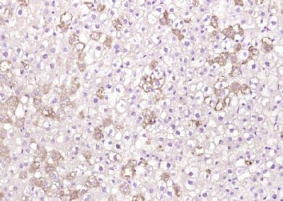
ApoM Polyclonal Antibody
Purified Rabbit Polyclonal Antibody (Pab)
- SPECIFICATION
- CITATIONS
- PROTOCOLS
- BACKGROUND

Application
| WB, IHC-P, IHC-F, IF, ICC, E |
|---|---|
| Primary Accession | O95445 |
| Reactivity | Rat, Pig, Dog, Bovine |
| Host | Rabbit |
| Clonality | Polyclonal |
| Calculated MW | 21 KDa |
| Physical State | Liquid |
| Immunogen | KLH conjugated synthetic peptide derived from human ApoM/Apolipoprotein M |
| Epitope Specificity | 101-188/188 |
| Isotype | IgG |
| Purity | affinity purified by Protein A |
| Buffer | 0.01M TBS (pH7.4) with 1% BSA, 0.02% Proclin300 and 50% Glycerol. |
| SUBCELLULAR LOCATION | Secreted. Present in high density lipoprotein (HDL) and to a lesser extent in triglyceride-rich lipoproteins (TGRLP) and low density lipoproteins. |
| SIMILARITY | Belongs to the calycin superfamily. Lipocalin family. Highly divergent. |
| Important Note | This product as supplied is intended for research use only, not for use in human, therapeutic or diagnostic applications. |
| Background Descriptions | Apolipoproteins are protein components of plasma lipoproteins. ApoM (Apolipoprotein M), also known as protein G3a, is a member of the Lipocalin family of proteins. ApoM is exclusively expressed in kidney tubular epithelial cells and liver hepatocytes. Mature ApoM retains its signal peptide, which acts as a hydrophobic anchor, and contains a structurally conserved eight stranded antiparallel ∫ barrel which binds retinol and retinoic acid. ApoM may play a key role in reverse cholesterol transport. It mainly associates with high density lipoprotein (HDL) and to a lesser extent with triglyceride-rich lipoprotein (TGRLP) and low-density lipoprotein (LDL). ApoM is important for the pre∫-HDL formation. Pre∫-HDL is an important acceptor of peripheral cellular cholesterol. The concentration of ApoM in plasma strongly correlates with total cholesterol. Low concentrations of ApoM in plasma is associated with diabetes. |
| Gene ID | 55937 |
|---|---|
| Other Names | Apolipoprotein M, Apo-M, ApoM, Protein G3a, APOM, G3A, NG20 |
| Target/Specificity | Plasma protein. Expressed in liver and kidney. |
| Dilution | WB=1:500-2000,IHC-P=1:100-500,IHC-F=1:100-500,ICC=1:100-500,IF=1:100-500,ELISA=1:5000-10000 |
| Storage | Store at -20 ℃ for one year. Avoid repeated freeze/thaw cycles. When reconstituted in sterile pH 7.4 0.01M PBS or diluent of antibody the antibody is stable for at least two weeks at 2-4 ℃. |
| Name | APOM |
|---|---|
| Synonyms | G3A, NG20 |
| Function | Probably involved in lipid transport. Can bind sphingosine-1- phosphate, myristic acid, palmitic acid and stearic acid, retinol, all- trans-retinoic acid and 9-cis-retinoic acid. |
| Cellular Location | Secreted. Note=Present in high density lipoprotein (HDL) and to a lesser extent in triglyceride-rich lipoproteins (TGRLP) and low density lipoproteins (LDL) |
| Tissue Location | Plasma protein. Expressed in liver and kidney. |
Research Areas
Citations (0)

Thousands of laboratories across the world have published research that depended on the performance of antibodies from Abcepta to advance their research. Check out links to articles that cite our products in major peer-reviewed journals, organized by research category.
Submit your citation using an Abcepta antibody to
info@abcepta.com, and receive a free "I Love Antibodies" mug.
info@abcepta.com, and receive a free "I Love Antibodies" mug.

Application Protocols
Provided below are standard protocols that you may find useful for product applications.
Abcepta welcomes feedback from its customers.
If you have used an Abcepta product and would like to share how it has performed, please click on the "Submit Review" button and provide the requested information. Our staff will examine and post your review and contact you if needed.
If you have any additional inquiries please email technical services at tech@abcepta.com.
$ 385.00
Cat# AP54867
Ordering Information
United States
AlbaniaAustraliaAustriaBelgiumBosnia & HerzegovinaBrazilBulgariaCanadaCentral AmericaChinaCroatiaCyprusCzech RepublicDenmarkEstoniaFinlandFranceGermanyGreeceHong KongHungaryIcelandIndiaIndonesiaIrelandIsraelItalyJapanLatviaLithuaniaLuxembourgMacedoniaMalaysiaMaltaMexicoNetherlandsNew ZealandNorwayPakistanPolandPortugalRomaniaSerbiaSingaporeSlovakiaSloveniaSouth AfricaSouth KoreaSpainSwedenSwitzerlandTaiwanTurkeyUnited KingdomUnited StatesVietnamWorldwideOthers
USA Headquarters
(888) 735-7227 / (858) 622-0099 or (858) 875-1900
Other Products
Shipping Information
Domestic orders (in stock items)
Shipped out the same day. Orders placed after 1 PM (PST) will ship out the next business day.
International orders
Contact your local distributors
