


 Foundational characteristics of cancer include proliferation, angiogenesis, migration, evasion of apoptosis, and cellular immortality. Find key markers for these cellular processes and antibodies to detect them.
 The SUMOplot™ Analysis Program predicts and scores sumoylation sites in your protein. SUMOylation is a post-translational modification involved in various cellular processes, such as nuclear-cytosolic transport, transcriptional regulation, apoptosis, protein stability, response to stress, and progression through the cell cycle.
 The Autophagy Receptor Motif Plotter predicts and scores autophagy receptor binding sites in your protein. Identifying proteins connected to this pathway is critical to understanding the role of autophagy in physiological as well as pathological processes such as development, differentiation, neurodegenerative diseases, stress, infection, and cancer.
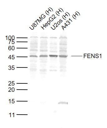
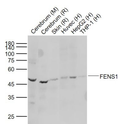
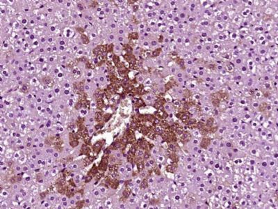
FENS1 Polyclonal Antibody
Purified Rabbit Polyclonal Antibody (Pab)
- SPECIFICATION
- CITATIONS
- PROTOCOLS
- BACKGROUND

Application
| WB, IHC-P, IHC-F, IF, ICC, E |
|---|---|
| Primary Accession | Q8IWB7 |
| Reactivity | Rat, Pig, Dog, Bovine |
| Host | Rabbit |
| Clonality | Polyclonal |
| Calculated MW | 46 KDa |
| Physical State | Liquid |
| Immunogen | KLH conjugated synthetic peptide derived from human FENS1 |
| Epitope Specificity | 21-120/410 |
| Isotype | IgG |
| Purity | affinity purified by Protein A |
| Buffer | 0.01M TBS (pH7.4) with 1% BSA, 0.02% Proclin300 and 50% Glycerol. |
| SUBCELLULAR LOCATION | Early endosome. |
| SIMILARITY | Contains 1 FYVE-type zinc finger. Contains 7 WD repeats. |
| SUBUNIT | Binds PtdIns3P in vitro with high specificity over other phosphoinositides. |
| Important Note | This product as supplied is intended for research use only, not for use in human, therapeutic or diagnostic applications. |
| Background Descriptions | WD-repeats are motifs that are found in a variety of proteins and are characterized by a conserved core of 40-60 amino acids that commonly form a tertiary propeller structure. While proteins that contain WD-repeats participate in a wide range of cellular functions, they are generally involved in regulatory mechanisms concerning chromatin assembly, cell cycle control, signal transduction, RNA processing, apoptosis and vesicular trafficking. WDFY1 (WD repeat and FYVE domain containing 1), also known as WDF1, FENS-1 or ZFYVE17, is a 410 amino acid protein that localizes to the early endosome and contains one FYVE-type zinc finger and seven WD repeats through which it may play a role in protein trafficking and signal transduction. |
| Gene ID | 57590 |
|---|---|
| Other Names | WD repeat and FYVE domain-containing protein 1, FYVE domain-containing protein localized to endosomes 1, FENS-1, Phosphoinositide-binding protein 1, WD40- and FYVE domain-containing protein 1, Zinc finger FYVE domain-containing protein 17, WDFY1 |
| Dilution | WB=1:500-2000,IHC-P=1:100-500,IHC-F=1:100-500,ICC=1:100-500,IF=1:100-500,ELISA=1:5000-10000 |
| Storage | Store at -20 ℃ for one year. Avoid repeated freeze/thaw cycles. When reconstituted in sterile pH 7.4 0.01M PBS or diluent of antibody the antibody is stable for at least two weeks at 2-4 ℃. |
| Name | WDFY1 |
|---|---|
| Function | Positively regulates TLR3- and TLR4-mediated signaling pathways by bridging the interaction between TLR3 or TLR4 and TICAM1. Promotes TLR3/4 ligand-induced activation of transcription factors IRF3 and NF-kappa-B, as well as the production of IFN-beta and inflammatory cytokines (PubMed:25736436). |
| Cellular Location | Early endosome |
Research Areas
Citations (0)

Thousands of laboratories across the world have published research that depended on the performance of antibodies from Abcepta to advance their research. Check out links to articles that cite our products in major peer-reviewed journals, organized by research category.
Submit your citation using an Abcepta antibody to
info@abcepta.com, and receive a free "I Love Antibodies" mug.
info@abcepta.com, and receive a free "I Love Antibodies" mug.

Application Protocols
Provided below are standard protocols that you may find useful for product applications.
Abcepta welcomes feedback from its customers.
If you have used an Abcepta product and would like to share how it has performed, please click on the "Submit Review" button and provide the requested information. Our staff will examine and post your review and contact you if needed.
If you have any additional inquiries please email technical services at tech@abcepta.com.
$ 385.00
Cat# AP55079
Ordering Information
United States
AlbaniaAustraliaAustriaBelgiumBosnia & HerzegovinaBrazilBulgariaCanadaCentral AmericaChinaCroatiaCyprusCzech RepublicDenmarkEstoniaFinlandFranceGermanyGreeceHong KongHungaryIcelandIndiaIndonesiaIrelandIsraelItalyJapanLatviaLithuaniaLuxembourgMacedoniaMalaysiaMaltaMexicoNetherlandsNew ZealandNorwayPakistanPolandPortugalRomaniaSerbiaSingaporeSlovakiaSloveniaSouth AfricaSouth KoreaSpainSwedenSwitzerlandTaiwanTurkeyUnited KingdomUnited StatesVietnamWorldwideOthers
USA Headquarters
(888) 735-7227 / (858) 622-0099 or (858) 875-1900
Other Products
Shipping Information
Domestic orders (in stock items)
Shipped out the same day. Orders placed after 1 PM (PST) will ship out the next business day.
International orders
Contact your local distributors
