


 Foundational characteristics of cancer include proliferation, angiogenesis, migration, evasion of apoptosis, and cellular immortality. Find key markers for these cellular processes and antibodies to detect them.
 The SUMOplot™ Analysis Program predicts and scores sumoylation sites in your protein. SUMOylation is a post-translational modification involved in various cellular processes, such as nuclear-cytosolic transport, transcriptional regulation, apoptosis, protein stability, response to stress, and progression through the cell cycle.
 The Autophagy Receptor Motif Plotter predicts and scores autophagy receptor binding sites in your protein. Identifying proteins connected to this pathway is critical to understanding the role of autophagy in physiological as well as pathological processes such as development, differentiation, neurodegenerative diseases, stress, infection, and cancer.
FUSIP1 Polyclonal Antibody
Purified Rabbit Polyclonal Antibody (Pab)
- SPECIFICATION
- CITATIONS
- PROTOCOLS
- BACKGROUND

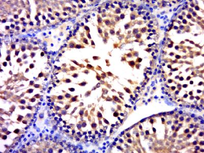
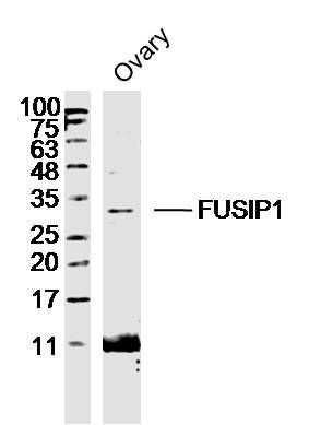
Application
| WB, IHC-P, IHC-F, IF, ICC, E |
|---|---|
| Primary Accession | O75494 |
| Reactivity | Rat, Pig, Dog, Bovine |
| Host | Rabbit |
| Clonality | Polyclonal |
| Calculated MW | 31 KDa |
| Physical State | Liquid |
| Immunogen | KLH conjugated synthetic peptide derived from human FUSIP1 |
| Epitope Specificity | 151-250/262 |
| Isotype | IgG |
| Purity | affinity purified by Protein A |
| Buffer | 0.01M TBS (pH7.4) with 1% BSA, 0.02% Proclin300 and 50% Glycerol. |
| SUBCELLULAR LOCATION | Nucleus speckle. Cytoplasm. |
| SIMILARITY | Belongs to the splicing factor SR family. Contains 1 RRM (RNA recognition motif) domain. |
| SUBUNIT | The phosphorylated but not the dephosphorylated form interacts with TRA2B/SFRS10. The dephosphorylated form interacts with SNRNP70. Isoform 1 and isoform 3 interact with FUS C-terminus. |
| Post-translational modifications | Phosphorylated. Fully dephosphorylated in mitosis and partially dephosphorylated on heat shock. Isoform 3 is phosphorylated on Ser-168. |
| Important Note | This product as supplied is intended for research use only, not for use in human, therapeutic or diagnostic applications. |
| Background Descriptions | FUSIP1 is a member of the Serine/Arginine (SR) family of splicing factors. Members of the SR family all contain one or more RNA recognition motifs (RRM) and an SR-rich domain. SR factors are not only essential for constitutive splicing but also regulate splicing in a concentration-dependent manner by influencing the selection of alternative splice sites. Expressed in a variety of tissues with low expression in kidney, liver and heart, FUSIP1 localizes to the cytoplasm and nuclear speckles. In its dephosphorylated form (occurring during M phase of the cell cycle), FUSIP1 functions as a potent general repressor of pre-mRNA splicing and can interact with U1 SnRNP 70. In its phosphorylated form, FUSIP1 interacts with Tra-2∫ and, together, they may cooperate in the regulation of splicing. Four isoforms exist for FUSIP1. In neurons, FUSIP1 isoforms may act to either positively or negatively regulate alternative splicing. |
| Gene ID | 10772 |
|---|---|
| Other Names | Serine/arginine-rich splicing factor 10, 40 kDa SR-repressor protein, SRrp40, FUS-interacting serine-arginine-rich protein 1, Splicing factor SRp38, Splicing factor, arginine/serine-rich 13A, TLS-associated protein with Ser-Arg repeats, TASR, TLS-associated protein with SR repeats, TLS-associated serine-arginine protein, TLS-associated SR protein, SRSF10, FUSIP1, FUSIP2, SFRS13A, TASR |
| Target/Specificity | Widely expressed. |
| Dilution | WB=1:500-2000,IHC-P=1:100-500,IHC-F=1:100-500,ICC=1:100-500,IF=1:100-500,ELISA=1:5000-10000 |
| Storage | Store at -20 ℃ for one year. Avoid repeated freeze/thaw cycles. When reconstituted in sterile pH 7.4 0.01M PBS or diluent of antibody the antibody is stable for at least two weeks at 2-4 ℃. |
| Name | SRSF10 |
|---|---|
| Synonyms | FUSIP1, FUSIP2, SFRS13A, TASR |
| Function | Splicing factor that in its dephosphorylated form acts as a general repressor of pre-mRNA splicing (PubMed:11684676, PubMed:12419250, PubMed:14765198). Seems to interfere with the U1 snRNP 5'-splice recognition of SNRNP70 (PubMed:14765198). Required for splicing repression in M-phase cells and after heat shock (PubMed:14765198). Also acts as a splicing factor that specifically promotes exon skipping during alternative splicing (PubMed:26876937). Interaction with YTHDC1, a RNA-binding protein that recognizes and binds N6-methyladenosine (m6A)-containing RNAs, prevents SRSF10 from binding to its mRNA-binding sites close to m6A-containing regions, leading to inhibit exon skipping during alternative splicing (PubMed:26876937). May be involved in regulation of alternative splicing in neurons, with isoform 1 acting as a positive and isoform 3 as a negative regulator (PubMed:12419250). |
| Cellular Location | Nucleus speckle. Cytoplasm |
| Tissue Location | Widely expressed. |
Citations (0)

Thousands of laboratories across the world have published research that depended on the performance of antibodies from Abcepta to advance their research. Check out links to articles that cite our products in major peer-reviewed journals, organized by research category.
Submit your citation using an Abcepta antibody to
info@abcepta.com, and receive a free "I Love Antibodies" mug.
info@abcepta.com, and receive a free "I Love Antibodies" mug.

Application Protocols
Provided below are standard protocols that you may find useful for product applications.
Abcepta welcomes feedback from its customers.
If you have used an Abcepta product and would like to share how it has performed, please click on the "Submit Review" button and provide the requested information. Our staff will examine and post your review and contact you if needed.
If you have any additional inquiries please email technical services at tech@abcepta.com.
$ 385.00
Cat# AP55099
Ordering Information
United States
AlbaniaAustraliaAustriaBelgiumBosnia & HerzegovinaBrazilBulgariaCanadaCentral AmericaChinaCroatiaCyprusCzech RepublicDenmarkEstoniaFinlandFranceGermanyGreeceHong KongHungaryIcelandIndiaIndonesiaIrelandIsraelItalyJapanLatviaLithuaniaLuxembourgMacedoniaMalaysiaMaltaMexicoNetherlandsNew ZealandNorwayPakistanPolandPortugalRomaniaSerbiaSingaporeSlovakiaSloveniaSouth AfricaSouth KoreaSpainSwedenSwitzerlandTaiwanTurkeyUnited KingdomUnited StatesVietnamWorldwideOthers
USA Headquarters
(888) 735-7227 / (858) 622-0099 or (858) 875-1900
Other Products
Shipping Information
Domestic orders (in stock items)
Shipped out the same day. Orders placed after 1 PM (PST) will ship out the next business day.
International orders
Contact your local distributors
