


 Foundational characteristics of cancer include proliferation, angiogenesis, migration, evasion of apoptosis, and cellular immortality. Find key markers for these cellular processes and antibodies to detect them.
 The SUMOplot™ Analysis Program predicts and scores sumoylation sites in your protein. SUMOylation is a post-translational modification involved in various cellular processes, such as nuclear-cytosolic transport, transcriptional regulation, apoptosis, protein stability, response to stress, and progression through the cell cycle.
 The Autophagy Receptor Motif Plotter predicts and scores autophagy receptor binding sites in your protein. Identifying proteins connected to this pathway is critical to understanding the role of autophagy in physiological as well as pathological processes such as development, differentiation, neurodegenerative diseases, stress, infection, and cancer.
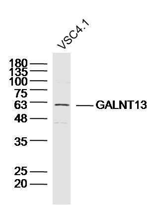
GALNT13 Polyclonal Antibody
Purified Rabbit Polyclonal Antibody (Pab)
- SPECIFICATION
- CITATIONS
- PROTOCOLS
- BACKGROUND

Application
| WB, IHC-P, IHC-F, IF, ICC, E |
|---|---|
| Primary Accession | Q8IUC8 |
| Reactivity | Rat, Pig, Dog, Bovine |
| Host | Rabbit |
| Clonality | Polyclonal |
| Calculated MW | 64 KDa |
| Physical State | Liquid |
| Immunogen | KLH conjugated synthetic peptide derived from human GALNT13/GalNAc-T13 |
| Epitope Specificity | 351-450/556 |
| Isotype | IgG |
| Purity | affinity purified by Protein A |
| Buffer | 0.01M TBS (pH7.4) with 1% BSA, 0.02% Proclin300 and 50% Glycerol. |
| SUBCELLULAR LOCATION | Golgi apparatus membrane; Single pass type II membrane protein. |
| SIMILARITY | Belongs to the glycosyltransferase 2 family. GalNAc-T subfamily. Contains 1 ricin B-type lectin domain. |
| Important Note | This product as supplied is intended for research use only, not for use in human, therapeutic or diagnostic applications. |
| Background Descriptions | The UDP-N-acetyl-alpha-D-galactosamine:polypeptide N-acetylgalactosaminyltransferase (GalNAc-T) family of enzymes are substrate-specific proteins that catalyze the transfer of GalNAc (N-acetylgalactosamine) to serine and threonine residues onto various proteins, thereby initiating mucin-type O-linked glycosylation in the Golgi apparatus. GalNAc-T13 (Polypeptide N-acetylgalactosaminyltransferase 13), also known as UDP-GalNAc:polypeptide N-acetylgalactosaminyltransferase 13, is a 556 amino acid protein that displays much stronger enzymatic activity than GalNAc-1 towards GalNAc transfer to mucin peptides such as Muc5a and Muc7. The N-terminal domain is involved in substrate binding and manganese coordination, while the C-terminal domain is involved in UDP-Gal binding and catalytic reaction. With specific expression in the central nervous system, GalNAc-T13 may be responsible for the synthesis of Tn antigen in neuronal cells, which is a universal carcinoma marker on malignant cells. |
| Gene ID | 114805 |
|---|---|
| Other Names | Polypeptide N-acetylgalactosaminyltransferase 13, 2.4.1.41, Polypeptide GalNAc transferase 13, GalNAc-T13, pp-GaNTase 13, Protein-UDP acetylgalactosaminyltransferase 13, UDP-GalNAc:polypeptide N-acetylgalactosaminyltransferase 13, GALNT13, KIAA1918 |
| Target/Specificity | Specifically expressed in neuronal cells. Expressed in fetal brain, whole adult brain, cerebral cortex and cerebellum. Not expressed in other tissues tested. |
| Dilution | WB=1:500-2000,IHC-P=1:100-500,IHC-F=1:100-500,ICC=1:100-500,IF=1:100-500,ELISA=1:5000-10000 |
| Storage | Store at -20 ℃ for one year. Avoid repeated freeze/thaw cycles. When reconstituted in sterile pH 7.4 0.01M PBS or diluent of antibody the antibody is stable for at least two weeks at 2-4 ℃. |
| Name | GALNT13 |
|---|---|
| Synonyms | KIAA1918 |
| Function | Catalyzes the initial reaction in O-linked oligosaccharide biosynthesis, the transfer of an N-acetyl-D-galactosamine (GalNAc) residue from UDP-GalNAc to a serine or threonine residue on the protein receptor (PubMed:12407114, PubMed:22186971). Generates GalNAc-O-Ser/Thr structure also known as Tn antigen, which itself is immunogenic but also serves as a precursor for the synthesis of different mucin-type O- glycan core structures (PubMed:12407114). Contributes to the synthesis of O-linked glycans on mucins and proteoglycans of the central nervous system. May promote neurogenesis through glycosylation and stabilization of PDPN (By similarity) (PubMed:12407114, PubMed:22186971). |
| Cellular Location | Golgi apparatus membrane; Single- pass type II membrane protein |
| Tissue Location | Specifically expressed in neuronal cells. Expressed in fetal brain, whole adult brain, cerebral cortex and cerebellum. Not expressed in other tissues tested. |
Citations (0)

Thousands of laboratories across the world have published research that depended on the performance of antibodies from Abcepta to advance their research. Check out links to articles that cite our products in major peer-reviewed journals, organized by research category.
Submit your citation using an Abcepta antibody to
info@abcepta.com, and receive a free "I Love Antibodies" mug.
info@abcepta.com, and receive a free "I Love Antibodies" mug.

Application Protocols
Provided below are standard protocols that you may find useful for product applications.
Abcepta welcomes feedback from its customers.
If you have used an Abcepta product and would like to share how it has performed, please click on the "Submit Review" button and provide the requested information. Our staff will examine and post your review and contact you if needed.
If you have any additional inquiries please email technical services at tech@abcepta.com.
$ 385.00
Cat# AP55117
Ordering Information
United States
AlbaniaAustraliaAustriaBelgiumBosnia & HerzegovinaBrazilBulgariaCanadaCentral AmericaChinaCroatiaCyprusCzech RepublicDenmarkEstoniaFinlandFranceGermanyGreeceHong KongHungaryIcelandIndiaIndonesiaIrelandIsraelItalyJapanLatviaLithuaniaLuxembourgMacedoniaMalaysiaMaltaMexicoNetherlandsNew ZealandNorwayPakistanPolandPortugalRomaniaSerbiaSingaporeSlovakiaSloveniaSouth AfricaSouth KoreaSpainSwedenSwitzerlandTaiwanTurkeyUnited KingdomUnited StatesVietnamWorldwideOthers
USA Headquarters
(888) 735-7227 / (858) 622-0099 or (858) 875-1900
Shipping Information
Domestic orders (in stock items)
Shipped out the same day. Orders placed after 1 PM (PST) will ship out the next business day.
International orders
Contact your local distributors
