


 Foundational characteristics of cancer include proliferation, angiogenesis, migration, evasion of apoptosis, and cellular immortality. Find key markers for these cellular processes and antibodies to detect them.
 The SUMOplot™ Analysis Program predicts and scores sumoylation sites in your protein. SUMOylation is a post-translational modification involved in various cellular processes, such as nuclear-cytosolic transport, transcriptional regulation, apoptosis, protein stability, response to stress, and progression through the cell cycle.
 The Autophagy Receptor Motif Plotter predicts and scores autophagy receptor binding sites in your protein. Identifying proteins connected to this pathway is critical to understanding the role of autophagy in physiological as well as pathological processes such as development, differentiation, neurodegenerative diseases, stress, infection, and cancer.
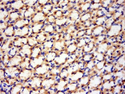
GLT28D1 Polyclonal Antibody
Purified Rabbit Polyclonal Antibody (Pab)
- SPECIFICATION
- CITATIONS
- PROTOCOLS
- BACKGROUND

Application
| WB, IHC-P, IHC-F, IF, ICC, E |
|---|---|
| Primary Accession | Q9NP73 |
| Reactivity | Rat |
| Host | Rabbit |
| Clonality | Polyclonal |
| Calculated MW | 126 KDa |
| Physical State | Liquid |
| Immunogen | KLH conjugated synthetic peptide derived from human GLT28D1/ALG13 |
| Epitope Specificity | 21-120/1137 |
| Isotype | IgG |
| Purity | affinity purified by Protein A |
| Buffer | 0.01M TBS (pH7.4) with 1% BSA, 0.02% Proclin300 and 50% Glycerol. |
| SUBCELLULAR LOCATION | Endoplasmic reticulum. Could be recruited to the cytosolic face of the endoplasmic reticulum membrane through its interaction with ALG14. |
| SIMILARITY | Belongs to the glycosyltransferase 28 family. Contains 1 OTU domain. Contains 1 Tudor domain. |
| SUBUNIT | Isoform 2 may interact with ALG14. |
| Important Note | This product as supplied is intended for research use only, not for use in human, therapeutic or diagnostic applications. |
| Background Descriptions | ALG13 is a 1,137 amino acid protein belonging to the glycosyltransferase 28 family. Encoded by a gene that maps to human chromosome Xq23, ALG13 is a subunit of a bipartite UDP-N-acetylglucosamine transferase and plays a role in protein folding regulation and stabilization. ALG13 contains one OTU domain, one TudorSN domain, and exists as four alternatively spliced isoforms. Heterodimerizing with ALG14, ALG13 forms a UDP-GlcNAc glycosyltransferase, which catalyzes the second sugar addition of the oligosaccharide precursor in endoplasmic reticulum (ER) N-linked glycosylation. ALG13 localizes to ER and may be recruited to the cytosolic face of the membrane by interacting with ALG14. |
| Gene ID | 79868 |
|---|---|
| Other Names | Putative bifunctional UDP-N-acetylglucosamine transferase and deubiquitinase ALG13, 2.4.1.141, 3.4.19.12, Asparagine-linked glycosylation 13 homolog, Glycosyltransferase 28 domain-containing protein 1, UDP-N-acetylglucosamine transferase subunit ALG13 homolog, ALG13, CXorf45, GLT28D1 |
| Dilution | WB=1:500-2000,IHC-P=1:100-500,IHC-F=1:100-500,ICC=1:100-500,IF=1:100-500,ELISA=1:5000-10000 |
| Storage | Store at -20 ℃ for one year. Avoid repeated freeze/thaw cycles. When reconstituted in sterile pH 7.4 0.01M PBS or diluent of antibody the antibody is stable for at least two weeks at 2-4 ℃. |
| Name | ALG13 (HGNC:30881) |
|---|---|
| Function | Catalytic subunit of the UDP-N-acetylglucosamine transferase complex that operates in the biosynthetic pathway of dolichol-linked oligosaccharides, the glycan precursors employed in protein asparagine (N)-glycosylation. The assembly of dolichol-linked oligosaccharides begins on the cytosolic side of the endoplasmic reticulum membrane and finishes in its lumen. The sequential addition of sugars to dolichol pyrophosphate produces dolichol-linked oligosaccharides containing fourteen sugars, including two GlcNAcs, nine mannoses and three glucoses. Once assembled, the oligosaccharide is transferred from the lipid to nascent proteins by oligosaccharyltransferases. On the cytoplasmic face of the endoplasmic reticulum, the dimeric ALG13/ALG14 complex catalyzes the second step of dolichol pyrophosphate biosynthesis, transferring a beta1,4-linked N-acetylglucosamine (GlcNAc) from UDP-GlcNAc to GlcNAc-pyrophosphatedolichol (Gn-PDol) to produce N,N'-diacetylchitobiosyl diphosphodolichol. N,N'- diacetylchitobiosyl diphosphodolichol is a substrate for ALG1, the following enzyme in the biosynthetic pathway. |
| Cellular Location | [Isoform 2]: Endoplasmic reticulum membrane; Peripheral membrane protein Note=Recruited to the cytosolic face of the endoplasmic reticulum membrane through its interaction with ALG14 |
Citations (0)

Thousands of laboratories across the world have published research that depended on the performance of antibodies from Abcepta to advance their research. Check out links to articles that cite our products in major peer-reviewed journals, organized by research category.
Submit your citation using an Abcepta antibody to
info@abcepta.com, and receive a free "I Love Antibodies" mug.
info@abcepta.com, and receive a free "I Love Antibodies" mug.

Application Protocols
Provided below are standard protocols that you may find useful for product applications.
Abcepta welcomes feedback from its customers.
If you have used an Abcepta product and would like to share how it has performed, please click on the "Submit Review" button and provide the requested information. Our staff will examine and post your review and contact you if needed.
If you have any additional inquiries please email technical services at tech@abcepta.com.
$ 385.00
Cat# AP55154
Ordering Information
United States
AlbaniaAustraliaAustriaBelgiumBosnia & HerzegovinaBrazilBulgariaCanadaCentral AmericaChinaCroatiaCyprusCzech RepublicDenmarkEstoniaFinlandFranceGermanyGreeceHong KongHungaryIcelandIndiaIndonesiaIrelandIsraelItalyJapanLatviaLithuaniaLuxembourgMacedoniaMalaysiaMaltaMexicoNetherlandsNew ZealandNorwayPakistanPolandPortugalRomaniaSerbiaSingaporeSlovakiaSloveniaSouth AfricaSouth KoreaSpainSwedenSwitzerlandTaiwanTurkeyUnited KingdomUnited StatesVietnamWorldwideOthers
USA Headquarters
(888) 735-7227 / (858) 622-0099 or (858) 875-1900
Shipping Information
Domestic orders (in stock items)
Shipped out the same day. Orders placed after 1 PM (PST) will ship out the next business day.
International orders
Contact your local distributors
