


 Foundational characteristics of cancer include proliferation, angiogenesis, migration, evasion of apoptosis, and cellular immortality. Find key markers for these cellular processes and antibodies to detect them.
 The SUMOplot™ Analysis Program predicts and scores sumoylation sites in your protein. SUMOylation is a post-translational modification involved in various cellular processes, such as nuclear-cytosolic transport, transcriptional regulation, apoptosis, protein stability, response to stress, and progression through the cell cycle.
 The Autophagy Receptor Motif Plotter predicts and scores autophagy receptor binding sites in your protein. Identifying proteins connected to this pathway is critical to understanding the role of autophagy in physiological as well as pathological processes such as development, differentiation, neurodegenerative diseases, stress, infection, and cancer.
GOSR1 Polyclonal Antibody
Purified Rabbit Polyclonal Antibody (Pab)
- SPECIFICATION
- CITATIONS
- PROTOCOLS
- BACKGROUND

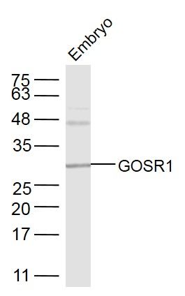
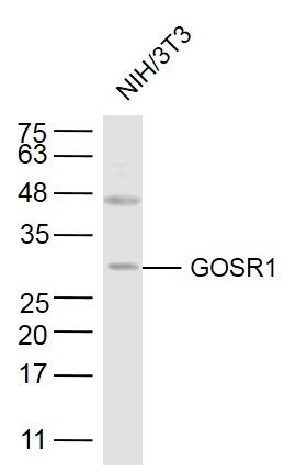
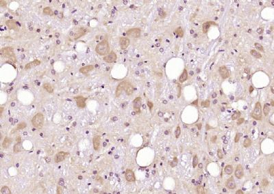
Application
| WB, IHC-P, IHC-F, IF, ICC, E |
|---|---|
| Primary Accession | O95249 |
| Reactivity | Rat |
| Host | Rabbit |
| Clonality | Polyclonal |
| Calculated MW | 29 KDa |
| Physical State | Liquid |
| Immunogen | KLH conjugated synthetic peptide derived from human GOSR1/GS28 |
| Epitope Specificity | 151-250/250 |
| Isotype | IgG |
| Purity | affinity purified by Protein A |
| Buffer | 0.01M TBS (pH7.4) with 1% BSA, 0.02% Proclin300 and 50% Glycerol. |
| SUBCELLULAR LOCATION | Cell Membrane, Endoplasmic reticulum and Golgi Apparatus |
| SIMILARITY | Belongs to the GOSR1 family. |
| SUBUNIT | Component of several multiprotein Golgi SNARE complexes. Identified in a SNARE complex with BET1, STX5 and YKT6, in a SNARE complex with BET1L, STX5 and YKT6, in a SNARE complex with STX5, GOSR2, SEC22B and BET1, and in complex with STX5 and COG3. Interacts with GABARAPL2 (By similarity). |
| Important Note | This product as supplied is intended for research use only, not for use in human, therapeutic or diagnostic applications. |
| Background Descriptions | In eukaryotic cells, the Golgi apparatus receives newly synthesized proteins from the endoplasmic reticulum and delivers them after covalent modification to their destination in the cell. For membrane-directed proteins this process is believed to be carried out via vesicular transport. Correct vesicular transport is determined by specific pairing of vesicle-associated SNAREs (v-SNAREs) with those on the target membrane (t-SNAREs). This complex then recruits soluble NSF attachment proteins (SNAPs) and N-ethylmaleimide-sensitive factor (NSF) to form the highly stable SNAP receptor (SNARE) complex. The formation of a SNARE complex pulls the vesicle and target membranes together and may provide the energy to drive the fusion of the lipid bilayers. Golgi SNARE 27 kDa (GS27) and GS28 belong to the SNARE protein family and are important trafficking proteins between the endoplasmic reticulum and the Golgi and between Golgi subcompartments. GS27 and GS28 both exist as cytoplasmically oriented integral membrane proteins. The human GS27 gene, which maps to chromosome 17q21, is located near a locus implicated in familial essential hypertension, indicating that it is a potential candidate gene for this disease. The human GS28 gene maps to chromosome 17q11. |
| Gene ID | 9527 |
|---|---|
| Other Names | Golgi SNAP receptor complex member 1, 28 kDa Golgi SNARE protein, 28 kDa cis-Golgi SNARE p28, GOS-28, GOSR1, GS28 |
| Dilution | WB=1:500-2000,IHC-P=1:100-500,IHC-F=1:100-500,ICC=1:100-500,IF=1:100-500,ELISA=1:5000-10000 |
| Storage | Store at -20 ℃ for one year. Avoid repeated freeze/thaw cycles. When reconstituted in sterile pH 7.4 0.01M PBS or diluent of antibody the antibody is stable for at least two weeks at 2-4 ℃. |
| Name | GOSR1 |
|---|---|
| Synonyms | GS28 |
| Function | Involved in transport from the ER to the Golgi apparatus as well as in intra-Golgi transport. It belongs to a super-family of proteins called t-SNAREs or soluble NSF (N-ethylmaleimide-sensitive factor) attachment protein receptor. May play a protective role against hydrogen peroxide induced cytotoxicity under glutathione depleted conditions in neuronal cells by regulating the intracellular ROS levels via inhibition of p38 MAPK (MAPK11, MAPK12, MAPK13 and MAPK14). Participates in docking and fusion stage of ER to cis-Golgi transport. Plays an important physiological role in VLDL-transport vesicle-Golgi fusion and thus in VLDL delivery to the hepatic cis-Golgi. |
| Cellular Location | Golgi apparatus membrane; Single-pass type IV membrane protein. Note=Localizes throughout the Golgi apparatus, with lowest levels in the trans-Golgi network (By similarity). Enriched on vesicular components at the terminal rims of the Golgi. Found in Golgi microtubules at low temperature (15 degrees Celsius). |
Research Areas
Citations (0)

Thousands of laboratories across the world have published research that depended on the performance of antibodies from Abcepta to advance their research. Check out links to articles that cite our products in major peer-reviewed journals, organized by research category.
Submit your citation using an Abcepta antibody to
info@abcepta.com, and receive a free "I Love Antibodies" mug.
info@abcepta.com, and receive a free "I Love Antibodies" mug.

Application Protocols
Provided below are standard protocols that you may find useful for product applications.
Abcepta welcomes feedback from its customers.
If you have used an Abcepta product and would like to share how it has performed, please click on the "Submit Review" button and provide the requested information. Our staff will examine and post your review and contact you if needed.
If you have any additional inquiries please email technical services at tech@abcepta.com.
$ 385.00
Cat# AP55181
Ordering Information
United States
AlbaniaAustraliaAustriaBelgiumBosnia & HerzegovinaBrazilBulgariaCanadaCentral AmericaChinaCroatiaCyprusCzech RepublicDenmarkEstoniaFinlandFranceGermanyGreeceHong KongHungaryIcelandIndiaIndonesiaIrelandIsraelItalyJapanLatviaLithuaniaLuxembourgMacedoniaMalaysiaMaltaMexicoNetherlandsNew ZealandNorwayPakistanPolandPortugalRomaniaSerbiaSingaporeSlovakiaSloveniaSouth AfricaSouth KoreaSpainSwedenSwitzerlandTaiwanTurkeyUnited KingdomUnited StatesVietnamWorldwideOthers
USA Headquarters
(888) 735-7227 / (858) 622-0099 or (858) 875-1900
Other Products
Shipping Information
Domestic orders (in stock items)
Shipped out the same day. Orders placed after 1 PM (PST) will ship out the next business day.
International orders
Contact your local distributors
