


 Foundational characteristics of cancer include proliferation, angiogenesis, migration, evasion of apoptosis, and cellular immortality. Find key markers for these cellular processes and antibodies to detect them.
 The SUMOplot™ Analysis Program predicts and scores sumoylation sites in your protein. SUMOylation is a post-translational modification involved in various cellular processes, such as nuclear-cytosolic transport, transcriptional regulation, apoptosis, protein stability, response to stress, and progression through the cell cycle.
 The Autophagy Receptor Motif Plotter predicts and scores autophagy receptor binding sites in your protein. Identifying proteins connected to this pathway is critical to understanding the role of autophagy in physiological as well as pathological processes such as development, differentiation, neurodegenerative diseases, stress, infection, and cancer.
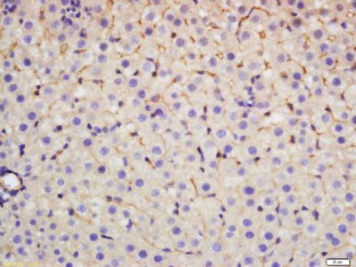
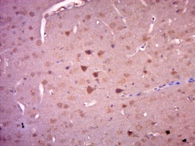
CCK39 Polyclonal Antibody
Purified Rabbit Polyclonal Antibody (Pab)
- SPECIFICATION
- CITATIONS
- PROTOCOLS
- BACKGROUND

Application
| IHC-P, IHC-F, IF, ICC, E |
|---|---|
| Primary Accession | P06307 |
| Reactivity | Rat, Pig, Dog, Bovine |
| Host | Rabbit |
| Clonality | Polyclonal |
| Calculated MW | 11 KDa |
| Physical State | Liquid |
| Immunogen | KLH conjugated synthetic peptide derived from human CCK39 |
| Epitope Specificity | 51-115/115 |
| Isotype | IgG |
| Purity | affinity purified by Protein A |
| Buffer | 0.01M TBS (pH7.4) with 1% BSA, 0.02% Proclin300 and 50% Glycerol. |
| SUBCELLULAR LOCATION | Secreted. |
| SIMILARITY | Belongs to the gastrin/cholecystokinin family. |
| SUBUNIT | Binds to CCK-A receptors in the pancreas and CCK-B receptors in the brain. |
| Post-translational modifications | The precursor is cleaved by proteases to produce a number of active cholecystokinins. |
| Important Note | This product as supplied is intended for research use only, not for use in human, therapeutic or diagnostic applications. |
| Background Descriptions | Cholecystokinin is a brain/gut peptide. In the gut, it induces the release of pancreatic enzymes and the contraction of the gallbladder. In the brain, its physiologic role is unclear. The cholecystokinin pro-hormone is processed by endo- and exo-proteolytic cleavages. Two transcript variants encoding the same protein have been found for this gene. [provided by RefSeq, Mar 2010]. |
| Gene ID | 885 |
|---|---|
| Other Names | Cholecystokinin, CCK, Cholecystokinin-58, CCK58, Cholecystokinin-58 desnonopeptide, (1-49)-CCK58, Cholecystokinin-39, CCK39, Cholecystokinin-33, CCK33, Cholecystokinin-25, CCK25, Cholecystokinin-18, CCK18, Cholecystokinin-12, CCK12, Cholecystokinin-8, CCK8, Cholecystokinin-7, CCK7, Cholecystokinin-5, CCK5, CCK |
| Target/Specificity | The shortest form (CCK8) is predominantly found in the brain, whereas the larger ones are found in the intestine. |
| Dilution | IHC-P=1:100-500,IHC-F=1:100-500,ICC=1:100-500,IF=1:100-500,ELISA=1:5000-10000 |
| Storage | Store at -20 ℃ for one year. Avoid repeated freeze/thaw cycles. When reconstituted in sterile pH 7.4 0.01M PBS or diluent of antibody the antibody is stable for at least two weeks at 2-4 ℃. |
| Name | CCK |
|---|---|
| Function | This peptide hormone induces gall bladder contraction and the release of pancreatic enzymes in the gut. Its function in the brain is not clear. Binding to CCK-A receptors stimulates amylase release from the pancreas, binding to CCK-B receptors stimulates gastric acid secretion. |
| Cellular Location | Secreted |
| Tissue Location | Detected in cerebrospinal fluid and urine (at protein level). |
Research Areas
Citations (0)

Thousands of laboratories across the world have published research that depended on the performance of antibodies from Abcepta to advance their research. Check out links to articles that cite our products in major peer-reviewed journals, organized by research category.
Submit your citation using an Abcepta antibody to
info@abcepta.com, and receive a free "I Love Antibodies" mug.
info@abcepta.com, and receive a free "I Love Antibodies" mug.

Application Protocols
Provided below are standard protocols that you may find useful for product applications.
Abcepta welcomes feedback from its customers.
If you have used an Abcepta product and would like to share how it has performed, please click on the "Submit Review" button and provide the requested information. Our staff will examine and post your review and contact you if needed.
If you have any additional inquiries please email technical services at tech@abcepta.com.
$ 385.00
Cat# AP55343
Ordering Information
United States
AlbaniaAustraliaAustriaBelgiumBosnia & HerzegovinaBrazilBulgariaCanadaCentral AmericaChinaCroatiaCyprusCzech RepublicDenmarkEstoniaFinlandFranceGermanyGreeceHong KongHungaryIcelandIndiaIndonesiaIrelandIsraelItalyJapanLatviaLithuaniaLuxembourgMacedoniaMalaysiaMaltaMexicoNetherlandsNew ZealandNorwayPakistanPolandPortugalRomaniaSerbiaSingaporeSlovakiaSloveniaSouth AfricaSouth KoreaSpainSwedenSwitzerlandTaiwanTurkeyUnited KingdomUnited StatesVietnamWorldwideOthers
USA Headquarters
(888) 735-7227 / (858) 622-0099 or (858) 875-1900
Shipping Information
Domestic orders (in stock items)
Shipped out the same day. Orders placed after 1 PM (PST) will ship out the next business day.
International orders
Contact your local distributors
