


 Foundational characteristics of cancer include proliferation, angiogenesis, migration, evasion of apoptosis, and cellular immortality. Find key markers for these cellular processes and antibodies to detect them.
 The SUMOplot™ Analysis Program predicts and scores sumoylation sites in your protein. SUMOylation is a post-translational modification involved in various cellular processes, such as nuclear-cytosolic transport, transcriptional regulation, apoptosis, protein stability, response to stress, and progression through the cell cycle.
 The Autophagy Receptor Motif Plotter predicts and scores autophagy receptor binding sites in your protein. Identifying proteins connected to this pathway is critical to understanding the role of autophagy in physiological as well as pathological processes such as development, differentiation, neurodegenerative diseases, stress, infection, and cancer.
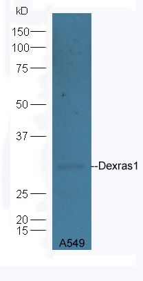
Dexras1 Polyclonal Antibody
Purified Rabbit Polyclonal Antibody (Pab)
- SPECIFICATION
- CITATIONS
- PROTOCOLS
- BACKGROUND

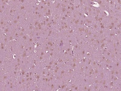
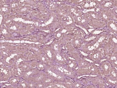
Application
| WB, IHC-P, IHC-F, IF, ICC, E |
|---|---|
| Primary Accession | Q9Y272 |
| Reactivity | Rat, Pig, Dog, Bovine |
| Host | Rabbit |
| Clonality | Polyclonal |
| Calculated MW | 31 KDa |
| Physical State | Liquid |
| Immunogen | KLH conjugated synthetic peptide derived from human Dexras1 |
| Epitope Specificity | 21-120/281 |
| Isotype | IgG |
| Purity | affinity purified by Protein A |
| Buffer | 0.01M TBS (pH7.4) with 1% BSA, 0.02% Proclin300 and 50% Glycerol. |
| SUBCELLULAR LOCATION | Cell membrane. Cytoplasm > perinuclear region. Nucleus. |
| SIMILARITY | Belongs to the small GTPase superfamily. RasD family. |
| Post-translational modifications | S-nitrosylation stimulates guanine-nucleotide exchange activity. |
| Important Note | This product as supplied is intended for research use only, not for use in human, therapeutic or diagnostic applications. |
| Background Descriptions | This gene encodes a member of the Ras superfamily of small GTPases and is induced by dexamethasone. The encoded protein is an activator of G-protein signaling and acts as a direct nucleotide exchange factor for Gi-Go proteins. This protein interacts with the neuronal nitric oxide adaptor protein CAPON, and a nuclear adaptor protein FE65, which interacts with the Alzheimer's disease amyloid precursor protein. This gene may play a role in dexamethasone-induced alterations in cell morphology, growth and cell-extracellular matrix interactions. Epigenetic inactivation of this gene is closely correlated with resistance to dexamethasone in multiple myeloma cells. Alternatively spliced transcript variants encoding different isoforms have been found for this gene.[provided by RefSeq, Sep 2011] |
| Gene ID | 51655 |
|---|---|
| Other Names | Dexamethasone-induced Ras-related protein 1, Activator of G-protein signaling 1, RASD1, AGS1, DEXRAS1 |
| Target/Specificity | Expressed in a variety of tissues including heart, cardiovascular tissues, brain, placenta, lung, liver, skeletal muscle, kidney, pancreas, gastrointestinal and reproductive tissues. |
| Dilution | WB=1:500-2000,IHC-P=1:100-500,IHC-F=1:100-500,ICC=1:100-500,IF=1:100-500,ELISA=1:5000-10000 |
| Storage | Store at -20 ℃ for one year. Avoid repeated freeze/thaw cycles. When reconstituted in sterile pH 7.4 0.01M PBS or diluent of antibody the antibody is stable for at least two weeks at 2-4 ℃. |
| Name | RASD1 |
|---|---|
| Synonyms | AGS1, DEXRAS1 |
| Function | Small GTPase. Negatively regulates the transcription regulation activity of the APBB1/FE65-APP complex via its interaction with APBB1/FE65 (By similarity). |
| Cellular Location | Cell membrane; Lipid-anchor; Cytoplasmic side. Cytoplasm, perinuclear region. Nucleus |
| Tissue Location | Expressed in a variety of tissues including heart, cardiovascular tissues, brain, placenta, lung, liver, skeletal muscle, kidney, pancreas, gastrointestinal and reproductive tissues |
Research Areas
Citations (0)

Thousands of laboratories across the world have published research that depended on the performance of antibodies from Abcepta to advance their research. Check out links to articles that cite our products in major peer-reviewed journals, organized by research category.
Submit your citation using an Abcepta antibody to
info@abcepta.com, and receive a free "I Love Antibodies" mug.
info@abcepta.com, and receive a free "I Love Antibodies" mug.

Application Protocols
Provided below are standard protocols that you may find useful for product applications.
Abcepta welcomes feedback from its customers.
If you have used an Abcepta product and would like to share how it has performed, please click on the "Submit Review" button and provide the requested information. Our staff will examine and post your review and contact you if needed.
If you have any additional inquiries please email technical services at tech@abcepta.com.
$ 385.00
Cat# AP55506
Ordering Information
United States
AlbaniaAustraliaAustriaBelgiumBosnia & HerzegovinaBrazilBulgariaCanadaCentral AmericaChinaCroatiaCyprusCzech RepublicDenmarkEstoniaFinlandFranceGermanyGreeceHong KongHungaryIcelandIndiaIndonesiaIrelandIsraelItalyJapanLatviaLithuaniaLuxembourgMacedoniaMalaysiaMaltaMexicoNetherlandsNew ZealandNorwayPakistanPolandPortugalRomaniaSerbiaSingaporeSlovakiaSloveniaSouth AfricaSouth KoreaSpainSwedenSwitzerlandTaiwanTurkeyUnited KingdomUnited StatesVietnamWorldwideOthers
USA Headquarters
(888) 735-7227 / (858) 622-0099 or (858) 875-1900
Shipping Information
Domestic orders (in stock items)
Shipped out the same day. Orders placed after 1 PM (PST) will ship out the next business day.
International orders
Contact your local distributors
