


 Foundational characteristics of cancer include proliferation, angiogenesis, migration, evasion of apoptosis, and cellular immortality. Find key markers for these cellular processes and antibodies to detect them.
 The SUMOplot™ Analysis Program predicts and scores sumoylation sites in your protein. SUMOylation is a post-translational modification involved in various cellular processes, such as nuclear-cytosolic transport, transcriptional regulation, apoptosis, protein stability, response to stress, and progression through the cell cycle.
 The Autophagy Receptor Motif Plotter predicts and scores autophagy receptor binding sites in your protein. Identifying proteins connected to this pathway is critical to understanding the role of autophagy in physiological as well as pathological processes such as development, differentiation, neurodegenerative diseases, stress, infection, and cancer.
FAM38A Polyclonal Antibody
Purified Rabbit Polyclonal Antibody (Pab)
- SPECIFICATION
- CITATIONS
- PROTOCOLS
- BACKGROUND

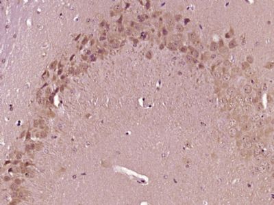
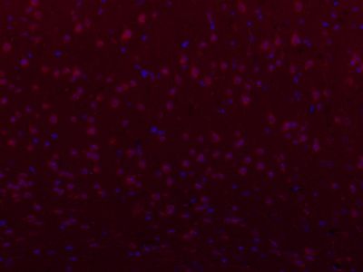
Application
| IHC-P, IHC-F, IF, ICC, E |
|---|---|
| Primary Accession | Q92508 |
| Reactivity | Rat, Dog, Bovine |
| Host | Rabbit |
| Clonality | Polyclonal |
| Calculated MW | 286790 Da |
| Gene ID | 9780 |
|---|---|
| Other Names | Piezo-type mechanosensitive ion channel component 1, Membrane protein induced by beta-amyloid treatment, Mib, Protein FAM38A, PIEZO1, FAM38A, KIAA0233 |
| Dilution | IHC-P=1:100-500,IHC-F=1:100-500,ICC=1:100,IF=1:100-500,Flow-Cyt=1ug/Test,ELISA=1:5000-10000 |
| Format | 0.01M TBS(pH7.4), 0.09% (W/V) sodium azide and 50% Glyce |
| Storage | Store at -20 ℃ for one year. Avoid repeated freeze/thaw cycles. When reconstituted in sterile pH 7.4 0.01M PBS or diluent of antibody the antibody is stable for at least two weeks at 2-4 ℃. |
| Name | PIEZO1 (HGNC:28993) |
|---|---|
| Synonyms | FAM38A, KIAA0233 |
| Function | Pore-forming subunit of the mechanosensitive non-specific cation Piezo channel required for rapidly adapting mechanically activated (MA) currents and has a key role in sensing touch and tactile pain (PubMed:23479567, PubMed:23695678, PubMed:25955826, PubMed:37590348). Piezo channels are homotrimeric three-blade propeller-shaped structures that utilize a cap-motion and plug-and- latch mechanism to gate their ion-conducting pathways (PubMed:37590348). Generates currents characterized by a linear current-voltage relationship that are sensitive to ruthenium red and gadolinium (By similarity). Conductance to monovalent alkali ions is highest for K(+), intermediate for Na(+) and lowest for Li(+) (PubMed:25955826). Divalent ions except for Mn(2+) permeate the channel but more slowly than the monovalent ions and they also reduce K(+) currents (PubMed:25955826). Plays a key role in epithelial cell adhesion by maintaining integrin activation through R-Ras recruitment to the ER, most probably in its activated state, and subsequent stimulation of calpain signaling (PubMed:20016066). In inner ear hair cells, PIEZO1/2 subunits may constitute part of the mechanotransducer (MET) non-selective cation channel complex where they may act as pore- forming ion-conducting component in the complex (By similarity). In the kidney, may contribute to the detection of intraluminal pressure changes and to urine flow sensing (By similarity). Acts as a shear- stress sensor that promotes endothelial cell organization and alignment in the direction of blood flow through calpain activation (PubMed:25119035). Plays a key role in blood vessel formation and vascular structure in both development and adult physiology (By similarity). Acts as a sensor of phosphatidylserine (PS) flipping at the plasma membrane and governs morphogenesis of muscle cells (By similarity). In myoblasts, flippase-mediated PS enrichment at the inner leaflet of plasma membrane triggers channel activation and Ca2+ influx followed by Rho GTPases signal transduction, leading to assembly of cortical actomyosin fibers and myotube formation (PubMed:29799007). |
| Cellular Location | Endoplasmic reticulum membrane; Multi-pass membrane protein {ECO:0000250|UniProtKB:E2JF22}. Endoplasmic reticulum-Golgi intermediate compartment membrane {ECO:0000250|UniProtKB:Q0KL00}. Cell membrane; Multi-pass membrane protein {ECO:0000250|UniProtKB:E2JF22}. Cell projection, lamellipodium membrane Note=In erythrocytes, located in the plasma membrane (PubMed:22529292, PubMed:23479567). Accumulates at the leading apical lamellipodia of endothelial cells in response to shear stress (PubMed:25119035) Colocalizes with F-actin and MYH9 at the actomyosin cortex in myoblasts. {ECO:0000250|UniProtKB:E2JF22, ECO:0000269|PubMed:22529292, ECO:0000269|PubMed:23479567, ECO:0000269|PubMed:25119035} |
| Tissue Location | Expressed in numerous tissues. In normal brain, expressed exclusively in neurons, not in astrocytes. In Alzheimer disease brains, expressed in about half of the activated astrocytes located around classical senile plaques. In Parkinson disease substantia nigra, not detected in melanin-containing neurons nor in activated astrocytes. Expressed in erythrocytes (at protein level) Expressed in myoblasts (at protein level) |
Citations (0)

Thousands of laboratories across the world have published research that depended on the performance of antibodies from Abcepta to advance their research. Check out links to articles that cite our products in major peer-reviewed journals, organized by research category.
Submit your citation using an Abcepta antibody to
info@abcepta.com, and receive a free "I Love Antibodies" mug.
info@abcepta.com, and receive a free "I Love Antibodies" mug.

Application Protocols
Provided below are standard protocols that you may find useful for product applications.
Abcepta welcomes feedback from its customers.
If you have used an Abcepta product and would like to share how it has performed, please click on the "Submit Review" button and provide the requested information. Our staff will examine and post your review and contact you if needed.
If you have any additional inquiries please email technical services at tech@abcepta.com.
$ 385.00
Cat# AP55728
Ordering Information
United States
AlbaniaAustraliaAustriaBelgiumBosnia & HerzegovinaBrazilBulgariaCanadaCentral AmericaChinaCroatiaCyprusCzech RepublicDenmarkEstoniaFinlandFranceGermanyGreeceHong KongHungaryIcelandIndiaIndonesiaIrelandIsraelItalyJapanLatviaLithuaniaLuxembourgMacedoniaMalaysiaMaltaMexicoNetherlandsNew ZealandNorwayPakistanPolandPortugalRomaniaSerbiaSingaporeSlovakiaSloveniaSouth AfricaSouth KoreaSpainSwedenSwitzerlandTaiwanTurkeyUnited KingdomUnited StatesVietnamWorldwideOthers
USA Headquarters
(888) 735-7227 / (858) 622-0099 or (858) 875-1900
Other Products
Shipping Information
Domestic orders (in stock items)
Shipped out the same day. Orders placed after 1 PM (PST) will ship out the next business day.
International orders
Contact your local distributors
