


 Foundational characteristics of cancer include proliferation, angiogenesis, migration, evasion of apoptosis, and cellular immortality. Find key markers for these cellular processes and antibodies to detect them.
 The SUMOplot™ Analysis Program predicts and scores sumoylation sites in your protein. SUMOylation is a post-translational modification involved in various cellular processes, such as nuclear-cytosolic transport, transcriptional regulation, apoptosis, protein stability, response to stress, and progression through the cell cycle.
 The Autophagy Receptor Motif Plotter predicts and scores autophagy receptor binding sites in your protein. Identifying proteins connected to this pathway is critical to understanding the role of autophagy in physiological as well as pathological processes such as development, differentiation, neurodegenerative diseases, stress, infection, and cancer.
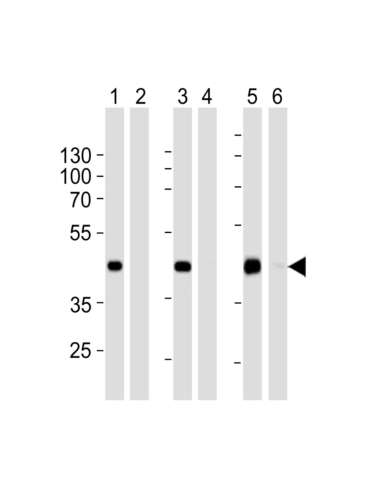
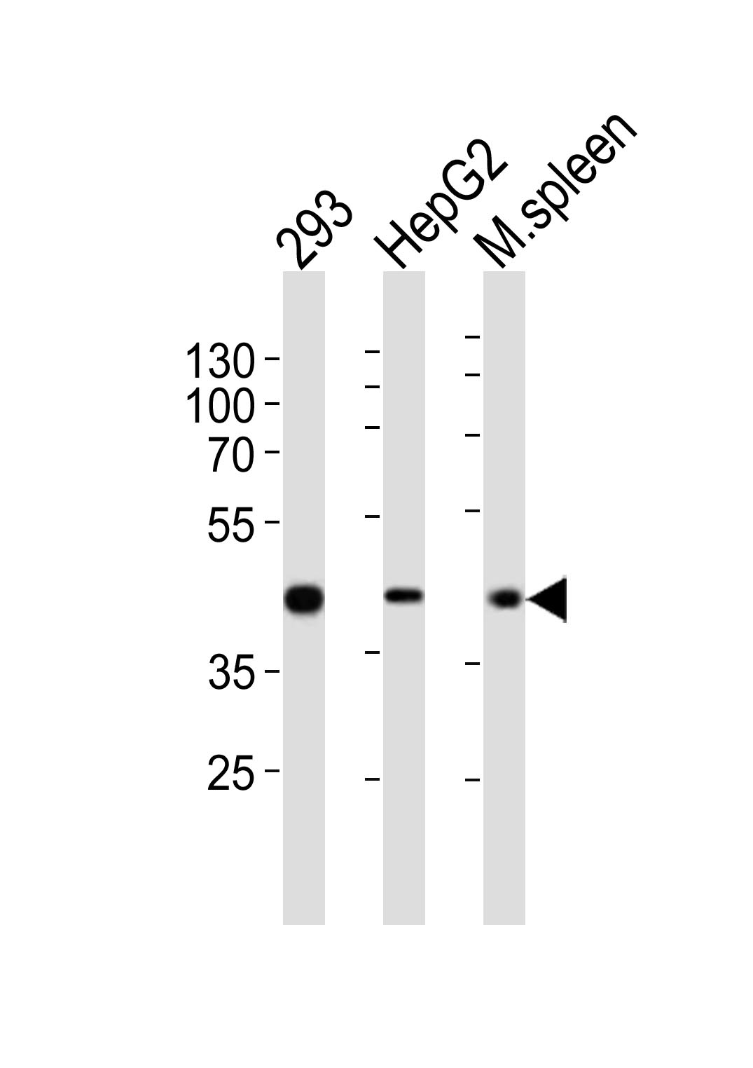
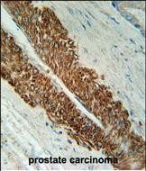
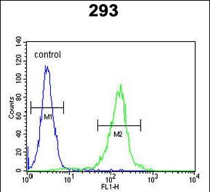
STEA2 Antibody (Center)
Affinity Purified Rabbit Polyclonal Antibody (Pab)
- SPECIFICATION
- CITATIONS
- PROTOCOLS
- BACKGROUND

Application
| WB, FC, IHC-P, E |
|---|---|
| Primary Accession | Q8NFT2 |
| Other Accession | NP_001035756.1 |
| Reactivity | Human, Mouse |
| Host | Rabbit |
| Clonality | Polyclonal |
| Isotype | Rabbit IgG |
| Calculated MW | 56056 Da |
| Antigen Region | 229-258 aa |
| Gene ID | 261729 |
|---|---|
| Other Names | Metalloreductase STEAP2, 1161-, Prostate cancer-associated protein 1, Protein up-regulated in metastatic prostate cancer, PUMPCn, Six-transmembrane epithelial antigen of prostate 2, SixTransMembrane protein of prostate 1, STEAP2, PCANAP1, STAMP1 |
| Target/Specificity | This STEAP2 antibody is generated from rabbits immunized with a KLH conjugated synthetic peptide between 229-258 amino acids of human STEAP2. |
| Dilution | WB~~1:500-1:1000 FC~~1:10~50 IHC-P~~1:50~100 E~~Use at an assay dependent concentration. |
| Format | Purified polyclonal antibody supplied in PBS with 0.09% (W/V) sodium azide. This antibody is purified through a protein A column, followed by peptide affinity purification. |
| Storage | Maintain refrigerated at 2-8°C for up to 2 weeks. For long term storage store at -20°C in small aliquots to prevent freeze-thaw cycles. |
| Precautions | STEA2 Antibody (Center) is for research use only and not for use in diagnostic or therapeutic procedures. |
| Name | STEAP2 |
|---|---|
| Synonyms | PCANAP1, STAMP1 |
| Function | Integral membrane protein that functions as a NADPH-dependent ferric-chelate reductase, using NADPH from one side of the membrane to reduce a Fe(3+) chelate that is bound on the other side of the membrane (By similarity). Mediates sequential transmembrane electron transfer from NADPH to FAD and onto heme, and finally to the Fe(3+) chelate (By similarity). Can also reduce Cu(2+) to Cu(1+) (By similarity). |
| Cellular Location | Endosome membrane {ECO:0000250|UniProtKB:Q8BWB6}; Multi-pass membrane protein. Cell membrane; Multi-pass membrane protein |
| Tissue Location | Expressed at high levels in prostate and at significantly lower levels in heart, brain, kidney, pancreas, and ovary. |

Thousands of laboratories across the world have published research that depended on the performance of antibodies from Abcepta to advance their research. Check out links to articles that cite our products in major peer-reviewed journals, organized by research category.
info@abcepta.com, and receive a free "I Love Antibodies" mug.

Provided below are standard protocols that you may find useful for product applications.
Background
This gene is a member of the STEAP family and encodes a multi-pass membrane protein that localizes to the Golgi complex, the plasma membrane, and the vesicular tubular structures in the cytosol. A highly similar protein in mouse has both ferrireductase and cupric reductase activity, and stimulates the cellular uptake of both iron and copper in vitro. Increased transcriptional expression of the human gene is associated with prostate cancer progression. Alternate transcriptional splice variants, encoding different isoforms, have been characterized.
References
Vaghjiani, R.J., et al. Tissue Eng Part A 15(8):2073-2083(2009)
Denoeud, F., et al. Genome Res. 17(6):746-759(2007)
Ohgami, R.S., et al. Blood 108(4):1388-1394(2006)
If you have used an Abcepta product and would like to share how it has performed, please click on the "Submit Review" button and provide the requested information. Our staff will examine and post your review and contact you if needed.
If you have any additional inquiries please email technical services at tech@abcepta.com.
Ordering Information
Other Products
Shipping Information
