


 Foundational characteristics of cancer include proliferation, angiogenesis, migration, evasion of apoptosis, and cellular immortality. Find key markers for these cellular processes and antibodies to detect them.
 The SUMOplot™ Analysis Program predicts and scores sumoylation sites in your protein. SUMOylation is a post-translational modification involved in various cellular processes, such as nuclear-cytosolic transport, transcriptional regulation, apoptosis, protein stability, response to stress, and progression through the cell cycle.
 The Autophagy Receptor Motif Plotter predicts and scores autophagy receptor binding sites in your protein. Identifying proteins connected to this pathway is critical to understanding the role of autophagy in physiological as well as pathological processes such as development, differentiation, neurodegenerative diseases, stress, infection, and cancer.
GPR91 Polyclonal Antibody
Purified Rabbit Polyclonal Antibody (Pab)
- SPECIFICATION
- CITATIONS
- PROTOCOLS
- BACKGROUND

Application
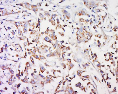
| WB, IHC-P, IHC-F, IF, ICC, E |
|---|---|
| Primary Accession | Q9BXA5 |
| Reactivity | Human, Mouse |
| Host | Rabbit |
| Clonality | Polyclonal |
| Calculated MW | 39 KDa |
| Physical State | Liquid |
| Immunogen | KLH conjugated synthetic peptide derived from human GPR91 |
| Epitope Specificity | 131-334/334 |
| Isotype | IgG |
| Purity | affinity purified by Protein A |
| Buffer | 0.01M TBS (pH7.4) with 1% BSA, 0.02% Proclin300 and 50% Glycerol. |
| SUBCELLULAR LOCATION | Cell membrane; Multi-pass membrane protein. |
| SIMILARITY | Belongs to the G-protein coupled receptor 1 family. |
| Important Note | This product as supplied is intended for research use only, not for use in human, therapeutic or diagnostic applications. |
| Background Descriptions | This gene encodes a G-protein-coupled receptor for succinate, an intermediate molecule of the citric acid cycle. It is involved in the promotion of hematopoietic progenitor cell development, and it has a potential role in renovascular hypertension which has known correlations to renal failure, diabetes and atherosclerosis. [provided by RefSeq, Oct 2009] |
| Gene ID | 56670 |
|---|---|
| Other Names | Succinate receptor 1, G-protein coupled receptor 91, P2Y purinoceptor 1-like, SUCNR1, GPR91 |
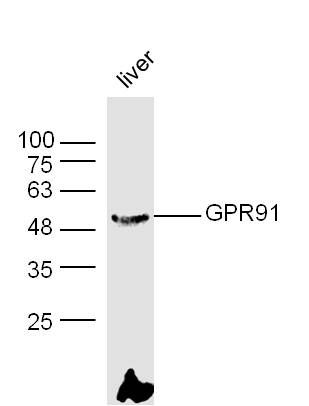
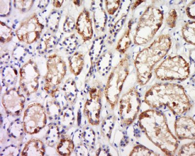
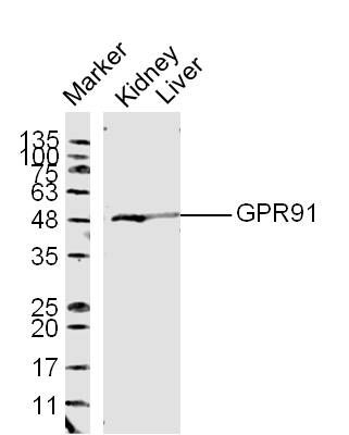
| Target/Specificity | Expressed specifically in kidney. |
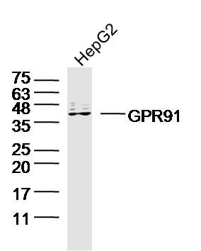
| Dilution | WB=1:500-2000,IHC-P=1:100-500,IHC-F=1:100-500,ICC=1:100-500,IF=1:100-500,ELISA=1:5000-10000 |
| Storage | Store at -20 ℃ for one year. Avoid repeated freeze/thaw cycles. When reconstituted in sterile pH 7.4 0.01M PBS or diluent of antibody the antibody is stable for at least two weeks at 2-4 ℃. |
| Name | SUCNR1 (HGNC:4542) |
|---|---|
| Synonyms | GPR91 |
| Function | G protein-coupled receptor for succinate able to mediate signaling through Gq/GNAQ or Gi/GNAI second messengers depending on the cell type and the processes regulated (By similarity) (PubMed:15141213, PubMed:23770096, PubMed:34133934). Succinate-SUCNR1 signaling serves as a link between metabolic stress, inflammation and energy homeostasis (PubMed:18820681, PubMed:34133934). In macrophages, plays a range of immune-regulatory roles. During inflammation, succinate-SUCNR1 signaling may act as an anti-inflammatory mediator or boost inflammation depending on the inflammatory status of cells (By similarity). Hyperpolarizes M2 macrophages versus M1 phenotype through Gq signaling by regulating the transcription of genes involved in immune function (PubMed:34133934). In activated M1 macrophages, plays a pro-inflammatory role in response to LPS (By similarity). Expressed in dendritic cells, where it is involved in the sensing of immunological danger and enhances immunity. Mediates succinate triggered intracelleular calcium mobilization, induces migratory responses and acts in synergy with Toll-like receptor ligands for the production of proinflammatory cytokines as well as an enhancement of antigen-specific activation of helper T cells (PubMed:18820681). In the small intestine, mediates the activation of tuft cells by dietary succinate and triggers type 2 immunity (By similarity). In adipocytes, plays an important role in the control of energy metabolism. In response to succinate, controls leptin expression in an AMPK-JNK-CEBPA-dependent as well as circadian clock-regulated manner (By similarity). In muscle tissue, is expressed in non-muscle cells and coordinates muscle remodeling in response to the succinate produced during exercise training in a paracrine manner (By similarity). In retina, acts as a mediator of vessel growth during retinal development. In response to succinate, regulates the production of angiogenic factors, including VEGF, by retinal ganglion neurons (By similarity). |
| Cellular Location | Cell membrane; Multi-pass membrane protein |
| Tissue Location | Expressed specifically in kidney (PubMed:11273702). Highly expressed in immature dendritic cells, expression rapidly downregulates after maturation. Also expressed in macrophages (PubMed:18820681). |
Research Areas
Citations (0)

Thousands of laboratories across the world have published research that depended on the performance of antibodies from Abcepta to advance their research. Check out links to articles that cite our products in major peer-reviewed journals, organized by research category.
Submit your citation using an Abcepta antibody to
info@abcepta.com, and receive a free "I Love Antibodies" mug.
info@abcepta.com, and receive a free "I Love Antibodies" mug.

Application Protocols
Provided below are standard protocols that you may find useful for product applications.
Abcepta welcomes feedback from its customers.
If you have used an Abcepta product and would like to share how it has performed, please click on the "Submit Review" button and provide the requested information. Our staff will examine and post your review and contact you if needed.
If you have any additional inquiries please email technical services at tech@abcepta.com.
$ 385.00
Cat# AP55977
Ordering Information
United States
AlbaniaAustraliaAustriaBelgiumBosnia & HerzegovinaBrazilBulgariaCanadaCentral AmericaChinaCroatiaCyprusCzech RepublicDenmarkEstoniaFinlandFranceGermanyGreeceHong KongHungaryIcelandIndiaIndonesiaIrelandIsraelItalyJapanLatviaLithuaniaLuxembourgMacedoniaMalaysiaMaltaMexicoNetherlandsNew ZealandNorwayPakistanPolandPortugalRomaniaSerbiaSingaporeSlovakiaSloveniaSouth AfricaSouth KoreaSpainSwedenSwitzerlandTaiwanTurkeyUnited KingdomUnited StatesVietnamWorldwideOthers
USA Headquarters
(888) 735-7227 / (858) 622-0099 or (858) 875-1900
Other Products
Shipping Information
Domestic orders (in stock items)
Shipped out the same day. Orders placed after 1 PM (PST) will ship out the next business day.
International orders
Contact your local distributors
