


 Foundational characteristics of cancer include proliferation, angiogenesis, migration, evasion of apoptosis, and cellular immortality. Find key markers for these cellular processes and antibodies to detect them.
 The SUMOplot™ Analysis Program predicts and scores sumoylation sites in your protein. SUMOylation is a post-translational modification involved in various cellular processes, such as nuclear-cytosolic transport, transcriptional regulation, apoptosis, protein stability, response to stress, and progression through the cell cycle.
 The Autophagy Receptor Motif Plotter predicts and scores autophagy receptor binding sites in your protein. Identifying proteins connected to this pathway is critical to understanding the role of autophagy in physiological as well as pathological processes such as development, differentiation, neurodegenerative diseases, stress, infection, and cancer.
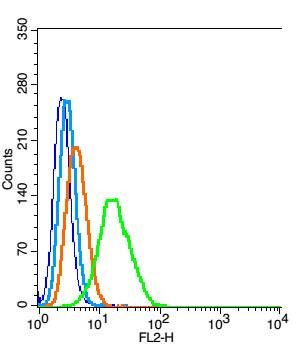
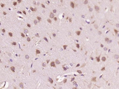
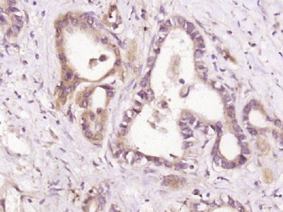
HDPTP Polyclonal Antibody
Purified Rabbit Polyclonal Antibody (Pab)
- SPECIFICATION
- CITATIONS
- PROTOCOLS
- BACKGROUND

Application
| IHC-P, IHC-F, IF, ICC, E |
|---|---|
| Primary Accession | Q9H3S7 |
| Reactivity | Rat, Pig |
| Host | Rabbit |
| Clonality | Polyclonal |
| Calculated MW | 179 KDa |
| Physical State | Liquid |
| Immunogen | KLH conjugated synthetic peptide derived from human HDPTP |
| Epitope Specificity | 1501-1636/1636 |
| Isotype | IgG |
| Purity | affinity purified by Protein A |
| Buffer | 0.01M TBS (pH7.4) with 1% BSA, 0.02% Proclin300 and 50% Glycerol. |
| SUBCELLULAR LOCATION | Nucleus. Cytoplasm. Cytoplasmic vesicle. Endosome. Cytoplasm, cytoskeleton, cilium basal body. |
| SIMILARITY | Belongs to the protein-tyrosine phosphatase family. Non-receptor class subfamily. Contains 1 BRO1 domain.Contains 2 TPR repeats.Contains 1 tyrosine-protein phosphatase domain. |
| SUBUNIT | Interacts with GRAP2 and GRB2. |
| Important Note | This product as supplied is intended for research use only, not for use in human, therapeutic or diagnostic applications. |
| Background Descriptions | HD-PTP is a 1,636 amino acid protein encoded by the human gene PTPN23. HD-PTP belongs to the protein-tyrosine phosphatase family, non-receptor class subfamily. It contains one BRO1 domain, two TPR repeats and one tyrosine-protein phosphatase domain. The C-terminal region contains the PTP-like domain, whereas the N-terminal region contains the two TPR regions. These regions are homologous to the yeast protein, BRO1, which is involved in the mitogen-activated protein kinase signaling pathway. Similarly, HD-PTP is believed to act as a negative regulator of Ras-mediated mitogenic activity and is phosphorylated upon DNA damage, probably by ATM or ATR. HD-PTP protein is differentially modulated by two angiogenic growth factors. While Vascular Endothelial Growth Factor (VEGF) has no affect on protein levels, Fibroblast Growth Factor-2 (FGF-2) induces HD-PTP degradation via the proteasome system. |
| Gene ID | 25930 |
|---|---|
| Other Names | Tyrosine-protein phosphatase non-receptor type 23, 3.1.3.48, His domain-containing protein tyrosine phosphatase, HD-PTP, Protein tyrosine phosphatase TD14, PTP-TD14, PTPN23, KIAA1471 |
| Dilution | IHC-P=1:100-500,IHC-F=1:100-500,ICC=1:100-500,IF=1:100-500,Flow-Cyt=1 µg/Test,ELISA=1:5000-10000 |
| Storage | Store at -20 ℃ for one year. Avoid repeated freeze/thaw cycles. When reconstituted in sterile pH 7.4 0.01M PBS or diluent of antibody the antibody is stable for at least two weeks at 2-4 ℃. |
| Name | PTPN23 |
|---|---|
| Synonyms | KIAA1471 |
| Function | Plays a role in sorting of endocytic ubiquitinated cargos into multivesicular bodies (MVBs) via its interaction with the ESCRT-I complex (endosomal sorting complex required for transport I), and possibly also other ESCRT complexes (PubMed:18434552, PubMed:21757351). May act as a negative regulator of Ras-mediated mitogenic activity (PubMed:18434552). Plays a role in ciliogenesis (PubMed:20393563). |
| Cellular Location | Nucleus. Cytoplasm. Cytoplasmic vesicle. Endosome. Cytoplasm, cytoskeleton, cilium basal body. Early endosome |
Research Areas
Citations (0)

Thousands of laboratories across the world have published research that depended on the performance of antibodies from Abcepta to advance their research. Check out links to articles that cite our products in major peer-reviewed journals, organized by research category.
Submit your citation using an Abcepta antibody to
info@abcepta.com, and receive a free "I Love Antibodies" mug.
info@abcepta.com, and receive a free "I Love Antibodies" mug.

Application Protocols
Provided below are standard protocols that you may find useful for product applications.
Abcepta welcomes feedback from its customers.
If you have used an Abcepta product and would like to share how it has performed, please click on the "Submit Review" button and provide the requested information. Our staff will examine and post your review and contact you if needed.
If you have any additional inquiries please email technical services at tech@abcepta.com.
$ 385.00
Cat# AP55991
Ordering Information
United States
AlbaniaAustraliaAustriaBelgiumBosnia & HerzegovinaBrazilBulgariaCanadaCentral AmericaChinaCroatiaCyprusCzech RepublicDenmarkEstoniaFinlandFranceGermanyGreeceHong KongHungaryIcelandIndiaIndonesiaIrelandIsraelItalyJapanLatviaLithuaniaLuxembourgMacedoniaMalaysiaMaltaMexicoNetherlandsNew ZealandNorwayPakistanPolandPortugalRomaniaSerbiaSingaporeSlovakiaSloveniaSouth AfricaSouth KoreaSpainSwedenSwitzerlandTaiwanTurkeyUnited KingdomUnited StatesVietnamWorldwideOthers
USA Headquarters
(888) 735-7227 / (858) 622-0099 or (858) 875-1900
Shipping Information
Domestic orders (in stock items)
Shipped out the same day. Orders placed after 1 PM (PST) will ship out the next business day.
International orders
Contact your local distributors
