


 Foundational characteristics of cancer include proliferation, angiogenesis, migration, evasion of apoptosis, and cellular immortality. Find key markers for these cellular processes and antibodies to detect them.
 The SUMOplot™ Analysis Program predicts and scores sumoylation sites in your protein. SUMOylation is a post-translational modification involved in various cellular processes, such as nuclear-cytosolic transport, transcriptional regulation, apoptosis, protein stability, response to stress, and progression through the cell cycle.
 The Autophagy Receptor Motif Plotter predicts and scores autophagy receptor binding sites in your protein. Identifying proteins connected to this pathway is critical to understanding the role of autophagy in physiological as well as pathological processes such as development, differentiation, neurodegenerative diseases, stress, infection, and cancer.
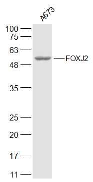
FOXJ2 Polyclonal Antibody
Purified Rabbit Polyclonal Antibody (Pab)
- SPECIFICATION
- CITATIONS
- PROTOCOLS
- BACKGROUND

Application
| WB, IHC-P, IHC-F, IF, ICC |
|---|---|
| Primary Accession | Q9P0K8 |
| Reactivity | Rat, Dog, Bovine |
| Host | Rabbit |
| Clonality | Polyclonal |
| Calculated MW | 62 KDa |
| Physical State | Liquid |
| Immunogen | KLH conjugated synthetic peptide derived from human FOXJ2 |
| Epitope Specificity | 111-210/574 |
| Isotype | IgG |
| Purity | affinity purified by Protein A |
| Buffer | 0.01M TBS (pH7.4) with 1% BSA, 0.02% Proclin300 and 50% Glycerol. |
| SUBCELLULAR LOCATION | Nucleus. |
| SIMILARITY | Contains 1 fork-head DNA-binding domain. |
| Post-translational modifications | Phosphorylated upon DNA damage, probably by ATM or ATR. |
| Important Note | This product as supplied is intended for research use only, not for use in human, therapeutic or diagnostic applications. |
| Background Descriptions | FOXJ2, Forkhead box protein J2, is a 574 amino acid protein encoded by the human gene FOXJ2. FOXJ2 is a novel forkhead factor, belonging to the forkhead family, with a dual DNA binding specificity. The HNF3/forkhead family includes a large number of transcription factors that share a structurally related DNA binding domain. forkhead factors are known to play important roles both during development and in adults. In the testis, FOXJ2 is expressed from pachytene spermatocytes to round spermatids, but not in spermatogonia. In addition to the germ lineage, only Sertoli cells of the testis showed expression of FOXJ2. In the ovary, only granulosa cells of the follicles express the factor. Neither mature spermatozoa nor oocytes have been found to express FOXJ2. However, both the trophectoderm (TE) and the inner cell mass (ICM) cell layers of the blastocyst express FOXJ2. |
| Gene ID | 55810 |
|---|---|
| Other Names | Forkhead box protein J2, Fork head homologous X, FOXJ2, FHX |
| Target/Specificity | Widely expressed. |
| Dilution | WB=1:500-2000,IHC-P=1:100-500,IHC-F=1:100-500,ICC=1:100-500,IF=1:100-500 |
| Storage | Store at -20 ℃ for one year. Avoid repeated freeze/thaw cycles. When reconstituted in sterile pH 7.4 0.01M PBS or diluent of antibody the antibody is stable for at least two weeks at 2-4 ℃. |
| Name | FOXJ2 |
|---|---|
| Synonyms | FHX |
| Function | [Isoform FOXJ2.L]: Transcriptional activator. Able to bind to two different type of DNA binding sites. More effective than isoform FOXJ2.S in transcriptional activation (PubMed:10777590, PubMed:10966786). Plays an important role in spermatogenesis, especially in spermatocyte meiosis (By similarity). |
| Cellular Location | Nucleus. |
| Tissue Location | Widely expressed. |
Citations (0)

Thousands of laboratories across the world have published research that depended on the performance of antibodies from Abcepta to advance their research. Check out links to articles that cite our products in major peer-reviewed journals, organized by research category.
Submit your citation using an Abcepta antibody to
info@abcepta.com, and receive a free "I Love Antibodies" mug.
info@abcepta.com, and receive a free "I Love Antibodies" mug.

Application Protocols
Provided below are standard protocols that you may find useful for product applications.
Abcepta welcomes feedback from its customers.
If you have used an Abcepta product and would like to share how it has performed, please click on the "Submit Review" button and provide the requested information. Our staff will examine and post your review and contact you if needed.
If you have any additional inquiries please email technical services at tech@abcepta.com.
$ 385.00
Cat# AP56162
Ordering Information
United States
AlbaniaAustraliaAustriaBelgiumBosnia & HerzegovinaBrazilBulgariaCanadaCentral AmericaChinaCroatiaCyprusCzech RepublicDenmarkEstoniaFinlandFranceGermanyGreeceHong KongHungaryIcelandIndiaIndonesiaIrelandIsraelItalyJapanLatviaLithuaniaLuxembourgMacedoniaMalaysiaMaltaMexicoNetherlandsNew ZealandNorwayPakistanPolandPortugalRomaniaSerbiaSingaporeSlovakiaSloveniaSouth AfricaSouth KoreaSpainSwedenSwitzerlandTaiwanTurkeyUnited KingdomUnited StatesVietnamWorldwideOthers
USA Headquarters
(888) 735-7227 / (858) 622-0099 or (858) 875-1900
Other Products
Shipping Information
Domestic orders (in stock items)
Shipped out the same day. Orders placed after 1 PM (PST) will ship out the next business day.
International orders
Contact your local distributors
