


 Foundational characteristics of cancer include proliferation, angiogenesis, migration, evasion of apoptosis, and cellular immortality. Find key markers for these cellular processes and antibodies to detect them.
 The SUMOplot™ Analysis Program predicts and scores sumoylation sites in your protein. SUMOylation is a post-translational modification involved in various cellular processes, such as nuclear-cytosolic transport, transcriptional regulation, apoptosis, protein stability, response to stress, and progression through the cell cycle.
 The Autophagy Receptor Motif Plotter predicts and scores autophagy receptor binding sites in your protein. Identifying proteins connected to this pathway is critical to understanding the role of autophagy in physiological as well as pathological processes such as development, differentiation, neurodegenerative diseases, stress, infection, and cancer.
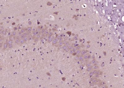
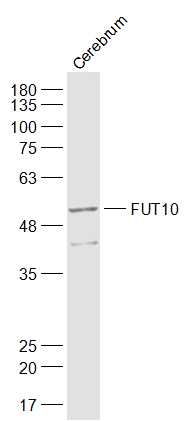
FUT10 Polyclonal Antibody
Purified Rabbit Polyclonal Antibody (Pab)

- SPECIFICATION
- CITATIONS
- PROTOCOLS
- BACKGROUND

Application
| WB, IHC-P, IHC-F, IF, ICC, E |
|---|---|
| Primary Accession | Q6P4F1 |
| Reactivity | Rat, Chimpanzee, Dog |
| Host | Rabbit |
| Clonality | Polyclonal |
| Calculated MW | 56 KDa |
| Physical State | Liquid |
| Immunogen | KLH conjugated synthetic peptide derived from human FUT10 |
| Epitope Specificity | 381-479/479 |
| Isotype | IgG |
| Purity | affinity purified by Protein A |
| Buffer | 0.01M TBS (pH7.4) with 1% BSA, 0.02% Proclin300 and 50% Glycerol. |
| SUBCELLULAR LOCATION | Golgi apparatus; Golgi stack membrane. |
| SIMILARITY | Belongs to the glycosyltransferase 10 family. |
| Important Note | This product as supplied is intended for research use only, not for use in human, therapeutic or diagnostic applications. |
| Background Descriptions | Glycosyltransferases that mediate the regio- and stereoselective transfer of sugars, such as the fucosyltransferases, determine cell surface-carbohydrate profiles, which is an essential interface for biological recognition processes. Fucosyltransferases catalyze the covalent association of fucose to different positional linkages in sugar acceptor molecules. Hematopoietic lineages rely on Fucosyltransferases to confer a surface carbohydrate phenotype, which mediates proper cell adhesion, molecule recruitment and cell trafficking. Localized to the Golgi apparatus as a single-pass transmembrane protein, FucT-X, also designated ?(1,3)-fucosyltransferase 10 or FUT10, is a 479 amino acid protein that is involved in protein modification and glycosylation. There are seven isoforms of FucT-X that are produced as a result of alternative splicing events. |
| Gene ID | 84750 |
|---|---|
| Other Names | Alpha-(1, 3)-fucosyltransferase 10, 2.4.1.-, Fucosyltransferase X, Fuc-TX, FucT-X, Galactoside 3-L-fucosyltransferase 10, Fucosyltransferase 10, FUT10 |
| Dilution | WB=1:500-2000,IHC-P=1:100-500,IHC-F=1:100-500,ICC=1:100-500,IF=1:100-500,ELISA=1:5000-10000 |
| Storage | Store at -20 ℃ for one year. Avoid repeated freeze/thaw cycles. When reconstituted in sterile pH 7.4 0.01M PBS or diluent of antibody the antibody is stable for at least two weeks at 2-4 ℃. |
| Name | POFUT3 {ECO:0000303|PubMed:39775168, ECO:0000312|HGNC:HGNC:19234} |
|---|---|
| Function | Protein O-fucosyltransferase that specifically catalyzes O- fucosylation of serine or threonine residues in EMI domains of target proteins, such as MMRN1, MMRN2 and EMID1 (PubMed:39775168). Attaches fucose through an O-glycosidic linkage (PubMed:39775168). O- fucosylation of EMI domain-containing proteins may be required for facilitating protein folding and secretion (PubMed:39775168). May also show alpha-(1,3)-fucosyltransferase activity toward the innermost N- acetyl glucosamine (GlcNAc) residue in biantennary N-glycan acceptors (PubMed:19088067). However, this was tested with a library of synthetic substrates and this activity is unsure in vivo (PubMed:19088067). May be involved in biosynthesis of Lewis X-carrying biantennary N-glycans that regulate neuron stem cell self-renewal during brain development (By similarity). |
| Cellular Location | Endoplasmic reticulum membrane; Single-pass type II membrane protein [Isoform 4]: Golgi apparatus. Lysosome |
| Tissue Location | Expressed in lung, digestive tract, gall bladder, placenta, kidney, uterus and brain. Not detected in spleen, heart, muscle, liver and pancreas. |
Research Areas
Citations (0)

Thousands of laboratories across the world have published research that depended on the performance of antibodies from Abcepta to advance their research. Check out links to articles that cite our products in major peer-reviewed journals, organized by research category.
Submit your citation using an Abcepta antibody to
info@abcepta.com, and receive a free "I Love Antibodies" mug.
info@abcepta.com, and receive a free "I Love Antibodies" mug.

Application Protocols
Provided below are standard protocols that you may find useful for product applications.
Abcepta welcomes feedback from its customers.
If you have used an Abcepta product and would like to share how it has performed, please click on the "Submit Review" button and provide the requested information. Our staff will examine and post your review and contact you if needed.
If you have any additional inquiries please email technical services at tech@abcepta.com.
$ 385.00
Cat# AP56174
Ordering Information
United States
AlbaniaAustraliaAustriaBelgiumBosnia & HerzegovinaBrazilBulgariaCanadaCentral AmericaChinaCroatiaCyprusCzech RepublicDenmarkEstoniaFinlandFranceGermanyGreeceHong KongHungaryIcelandIndiaIndonesiaIrelandIsraelItalyJapanLatviaLithuaniaLuxembourgMacedoniaMalaysiaMaltaMexicoNetherlandsNew ZealandNorwayPakistanPolandPortugalRomaniaSerbiaSingaporeSlovakiaSloveniaSouth AfricaSouth KoreaSpainSwedenSwitzerlandTaiwanTurkeyUnited KingdomUnited StatesVietnamWorldwideOthers
USA Headquarters
(888) 735-7227 / (858) 622-0099 or (858) 875-1900
Shipping Information
Domestic orders (in stock items)
Shipped out the same day. Orders placed after 1 PM (PST) will ship out the next business day.
International orders
Contact your local distributors
