


 Foundational characteristics of cancer include proliferation, angiogenesis, migration, evasion of apoptosis, and cellular immortality. Find key markers for these cellular processes and antibodies to detect them.
 The SUMOplot™ Analysis Program predicts and scores sumoylation sites in your protein. SUMOylation is a post-translational modification involved in various cellular processes, such as nuclear-cytosolic transport, transcriptional regulation, apoptosis, protein stability, response to stress, and progression through the cell cycle.
 The Autophagy Receptor Motif Plotter predicts and scores autophagy receptor binding sites in your protein. Identifying proteins connected to this pathway is critical to understanding the role of autophagy in physiological as well as pathological processes such as development, differentiation, neurodegenerative diseases, stress, infection, and cancer.
KTEL1 Polyclonal Antibody
Purified Rabbit Polyclonal Antibody (Pab)
- SPECIFICATION
- CITATIONS
- PROTOCOLS
- BACKGROUND

Application
| WB, IHC-P, IHC-F, IF, ICC, E |
|---|---|
| Primary Accession | Q8NBL1 |
| Reactivity | Rat, Dog, Bovine |
| Host | Rabbit |
| Clonality | Polyclonal |
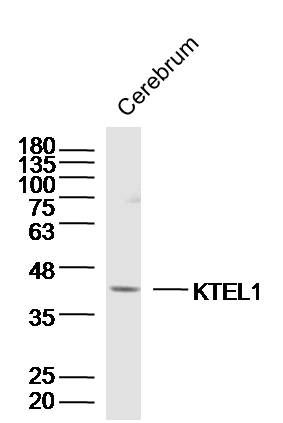
| Calculated MW | 43 KDa |
| Physical State | Liquid |
| Immunogen | KLH conjugated synthetic peptide derived from human KTEL1 |
| Epitope Specificity | 201-300/392 |
| Isotype | IgG |
| Purity | affinity purified by Protein A |
| Buffer | 0.01M TBS (pH7.4) with 1% BSA, 0.02% Proclin300 and 50% Glycerol. |
| SUBCELLULAR LOCATION | Endoplasmic reticulum lumen. |
| SIMILARITY | Belongs to the glycosyltransferase 90 family. |
| Important Note | This product as supplied is intended for research use only, not for use in human, therapeutic or diagnostic applications. |
| Background Descriptions | CLP46 is a KTEL motif-containing protein that belongs to the CAP10 family. The KTEL motif at the C-terminus of CLP46 is an endoplasmic reticulum (ER) retention signal which localizes the CLP46 protein to the lumen of the ER. CLP46 is thought to promote cell proliferation and is also believed to be involved with hepatic functions. The Drosophila protein rumi, a CAP10 protein and likely similar in function to CLP46, uses glucosylation to act as a Notch signaling regulator. Expressed in varying degrees in most adult tissue, CLP46 is especially abundant in the liver. CLP46 is not expressed in detectable levels in colon, thymus or small intestine. Human CLP46 shares 94% and 91% sequence similarity with its bovine and mouse homologs, respectively. |
| Gene ID | 56983 |
|---|---|
| Other Names | Protein O-glucosyltransferase 1, 2.4.1.376, CAP10-like 46 kDa protein, hCLP46, KTEL motif-containing protein 1, Myelodysplastic syndromes relative protein, O-glucosyltransferase Rumi homolog, hRumi, Protein O-xylosyltransferase POGLUT1, 2.4.2.63, POGLUT1 (HGNC:22954) |
| Target/Specificity | Expressed in most adult tissues at different intensities. Abundantly expressed in liver. Expressed also in brain, heart, skeletal muscle, spleen, kidney, placenta, lung and peripheral blood leukocyte. Not detectable in colon, thymus and small intestine. |
| Dilution | WB=1:500-2000,IHC-P=1:100-500,IHC-F=1:100-500,ICC=1:100-500,IF=1:100-500,ELISA=1:5000-10000 |
| Storage | Store at -20 ℃ for one year. Avoid repeated freeze/thaw cycles. When reconstituted in sterile pH 7.4 0.01M PBS or diluent of antibody the antibody is stable for at least two weeks at 2-4 ℃. |
| Name | POGLUT1 (HGNC:22954) |
|---|---|
| Function | Dual specificity glycosyltransferase that catalyzes the transfer of glucose and xylose from UDP-glucose and UDP-xylose, respectively, to a serine residue found in the consensus sequence of C- X-S-X-P-C (PubMed:21081508, PubMed:21490058, PubMed:21949356, PubMed:27807076, PubMed:28775322). Specifically targets extracellular EGF repeats of protein such as CRB2, F7, F9 and NOTCH2 (PubMed:21081508, PubMed:21490058, PubMed:21949356, PubMed:27807076, PubMed:28775322). Acts as a positive regulator of Notch signaling by mediating O-glucosylation of Notch, leading to regulate muscle development (PubMed:27807076). Notch glucosylation does not affect Notch ligand binding (PubMed:21490058). Required during early development to promote gastrulation: acts by mediating O-glucosylation of CRB2, which is required for CRB2 localization to the cell membrane (By similarity). |
| Cellular Location | Endoplasmic reticulum lumen |
| Tissue Location | Expressed in most adult tissues at different intensities. Abundantly expressed in liver. Expressed also in brain, heart, skeletal muscle, spleen, kidney, placenta, lung and peripheral blood leukocyte. Not detectable in colon, thymus and small intestine Expressed in the epidermis, especially in the upper parts, stratum spinosum and stratum granulosum (at protein level) |
Research Areas
Citations (0)

Thousands of laboratories across the world have published research that depended on the performance of antibodies from Abcepta to advance their research. Check out links to articles that cite our products in major peer-reviewed journals, organized by research category.
Submit your citation using an Abcepta antibody to
info@abcepta.com, and receive a free "I Love Antibodies" mug.
info@abcepta.com, and receive a free "I Love Antibodies" mug.

Application Protocols
Provided below are standard protocols that you may find useful for product applications.
Abcepta welcomes feedback from its customers.
If you have used an Abcepta product and would like to share how it has performed, please click on the "Submit Review" button and provide the requested information. Our staff will examine and post your review and contact you if needed.
If you have any additional inquiries please email technical services at tech@abcepta.com.
$ 385.00
Cat# AP56443
Ordering Information
United States
AlbaniaAustraliaAustriaBelgiumBosnia & HerzegovinaBrazilBulgariaCanadaCentral AmericaChinaCroatiaCyprusCzech RepublicDenmarkEstoniaFinlandFranceGermanyGreeceHong KongHungaryIcelandIndiaIndonesiaIrelandIsraelItalyJapanLatviaLithuaniaLuxembourgMacedoniaMalaysiaMaltaMexicoNetherlandsNew ZealandNorwayPakistanPolandPortugalRomaniaSerbiaSingaporeSlovakiaSloveniaSouth AfricaSouth KoreaSpainSwedenSwitzerlandTaiwanTurkeyUnited KingdomUnited StatesVietnamWorldwideOthers
USA Headquarters
(888) 735-7227 / (858) 622-0099 or (858) 875-1900
Shipping Information
Domestic orders (in stock items)
Shipped out the same day. Orders placed after 1 PM (PST) will ship out the next business day.
International orders
Contact your local distributors
