


 Foundational characteristics of cancer include proliferation, angiogenesis, migration, evasion of apoptosis, and cellular immortality. Find key markers for these cellular processes and antibodies to detect them.
 The SUMOplot™ Analysis Program predicts and scores sumoylation sites in your protein. SUMOylation is a post-translational modification involved in various cellular processes, such as nuclear-cytosolic transport, transcriptional regulation, apoptosis, protein stability, response to stress, and progression through the cell cycle.
 The Autophagy Receptor Motif Plotter predicts and scores autophagy receptor binding sites in your protein. Identifying proteins connected to this pathway is critical to understanding the role of autophagy in physiological as well as pathological processes such as development, differentiation, neurodegenerative diseases, stress, infection, and cancer.
Kindlin Polyclonal Antibody
Purified Rabbit Polyclonal Antibody (Pab)

- SPECIFICATION
- CITATIONS
- PROTOCOLS
- BACKGROUND

Application
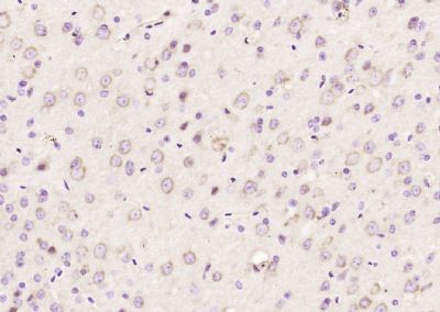
| IHC-P, IHC-F, IF, ICC, E |
|---|---|
| Primary Accession | Q9BQL6 |
| Reactivity | Rat, Pig, Dog |
| Host | Rabbit |
| Clonality | Polyclonal |
| Calculated MW | 77 KDa |
| Physical State | Liquid |
| Immunogen | KLH conjugated synthetic peptide derived from human Kindlin |
| Epitope Specificity | 601-677/677 |
| Isotype | IgG |
| Purity | affinity purified by Protein A |
| Buffer | 0.01M TBS (pH7.4) with 1% BSA, 0.02% Proclin300 and 50% Glycerol. |
| SUBCELLULAR LOCATION | Cytoplasm > cytoskeleton. Cell junction > focal adhesion. Cell projection > ruffle membrane. Constituent of focal adhesions. Localized at the basal aspect of skin keratinocytes, close to the cell membrane. Colocalizes with filamentous actin. Upon TGFB1 treatment, it localizes to membrane ruffles. |
| SIMILARITY | Belongs to the kindlin family. Contains 1 FERM domain. Contains 1 PH domain. |
| DISEASE | Defects in FERMT1 are the cause of Kindler syndrome (KINDS) [MIM:173650]. An autosomal recessive skin disorder characterized by skin blistering, photosensitivity, progressive poikiloderma, and extensive skin atrophy. Additional clinical features include gingival erosions, ocular, esophageal, gastrointestinal and urogenital involvement, and an increased risk of mucocutaneous malignancy. Note=Although most FERMT1 mutations are predicted to lead to premature termination of translation, and to loss of FERMT1 function, significant clinical variability is observed among patients. There is an association of FERMT1 missense and in-frame deletion mutations with milder disease phenotypes, and later onset of complications (PubMed:21936020). |
| Important Note | This product as supplied is intended for research use only, not for use in human, therapeutic or diagnostic applications. |
| Background Descriptions | This gene encodes a member of the fermitin family, and contains a FERM domain and a pleckstrin homology domain. The encoded protein is involved in integrin signaling and linkage of the actin cytoskeleton to the extracellular matrix. Mutations in this gene have been linked to Kindler syndrome. [provided by RefSeq, Dec 2009] |
| Gene ID | 55612 |
|---|---|
| Other Names | Fermitin family homolog 1, Kindlerin, Kindlin syndrome protein, Kindlin-1, Unc-112-related protein 1, FERMT1, C20orf42, KIND1, URP1 |
| Target/Specificity | Expressed in brain, skeletal muscle, kidney, colon, adrenal gland, prostate, and placenta. Weakly or not expressed in heart, thymus, spleen, liver, small intestine, bone marrow, lung and peripheral blood leukocytes. Overexpressed in some colon and lung tumors. In skin, it is localized within the epidermis and particularly in basal keratocytes. Not detected in epidermal melanocytes and dermal fibroblasts. |
| Dilution | IHC-P=1:100-500,IHC-F=1:100-500,ICC=1:100-500,IF=1:100-500,ELISA=1:5000-10000 |
| Storage | Store at -20 ℃ for one year. Avoid repeated freeze/thaw cycles. When reconstituted in sterile pH 7.4 0.01M PBS or diluent of antibody the antibody is stable for at least two weeks at 2-4 ℃. |
| Name | FERMT1 |
|---|---|
| Synonyms | C20orf42, KIND1, URP1 |
| Function | Involved in cell adhesion. Contributes to integrin activation. When coexpressed with talin, potentiates activation of ITGA2B. Required for normal keratinocyte proliferation. Required for normal polarization of basal keratinocytes in skin, and for normal cell shape. Required for normal adhesion of keratinocytes to fibronectin and laminin, and for normal keratinocyte migration to wound sites. May mediate TGF-beta 1 signaling in tumor progression. |
| Cellular Location | Cytoplasm, cytoskeleton. Cell junction, focal adhesion. Cell projection, ruffle membrane; Peripheral membrane protein; Cytoplasmic side. Note=Constituent of focal adhesions Localized at the basal aspect of skin keratinocytes, close to the cell membrane. Colocalizes with filamentous actin. Upon TGFB1 treatment, it localizes to membrane ruffles |
| Tissue Location | Expressed in brain, skeletal muscle, kidney, colon, adrenal gland, prostate, and placenta. Weakly or not expressed in heart, thymus, spleen, liver, small intestine, bone marrow, lung and peripheral blood leukocytes. Overexpressed in some colon and lung tumors. In skin, it is localized within the epidermis and particularly in basal keratocytes. Not detected in epidermal melanocytes and dermal fibroblasts. |
Citations (0)

Thousands of laboratories across the world have published research that depended on the performance of antibodies from Abcepta to advance their research. Check out links to articles that cite our products in major peer-reviewed journals, organized by research category.
Submit your citation using an Abcepta antibody to
info@abcepta.com, and receive a free "I Love Antibodies" mug.
info@abcepta.com, and receive a free "I Love Antibodies" mug.

Application Protocols
Provided below are standard protocols that you may find useful for product applications.
Abcepta welcomes feedback from its customers.
If you have used an Abcepta product and would like to share how it has performed, please click on the "Submit Review" button and provide the requested information. Our staff will examine and post your review and contact you if needed.
If you have any additional inquiries please email technical services at tech@abcepta.com.
$ 385.00
Cat# AP56543
Ordering Information
United States
AlbaniaAustraliaAustriaBelgiumBosnia & HerzegovinaBrazilBulgariaCanadaCentral AmericaChinaCroatiaCyprusCzech RepublicDenmarkEstoniaFinlandFranceGermanyGreeceHong KongHungaryIcelandIndiaIndonesiaIrelandIsraelItalyJapanLatviaLithuaniaLuxembourgMacedoniaMalaysiaMaltaMexicoNetherlandsNew ZealandNorwayPakistanPolandPortugalRomaniaSerbiaSingaporeSlovakiaSloveniaSouth AfricaSouth KoreaSpainSwedenSwitzerlandTaiwanTurkeyUnited KingdomUnited StatesVietnamWorldwideOthers
USA Headquarters
(888) 735-7227 / (858) 622-0099 or (858) 875-1900
Other Products
Shipping Information
Domestic orders (in stock items)
Shipped out the same day. Orders placed after 1 PM (PST) will ship out the next business day.
International orders
Contact your local distributors
