


 Foundational characteristics of cancer include proliferation, angiogenesis, migration, evasion of apoptosis, and cellular immortality. Find key markers for these cellular processes and antibodies to detect them.
 The SUMOplot™ Analysis Program predicts and scores sumoylation sites in your protein. SUMOylation is a post-translational modification involved in various cellular processes, such as nuclear-cytosolic transport, transcriptional regulation, apoptosis, protein stability, response to stress, and progression through the cell cycle.
 The Autophagy Receptor Motif Plotter predicts and scores autophagy receptor binding sites in your protein. Identifying proteins connected to this pathway is critical to understanding the role of autophagy in physiological as well as pathological processes such as development, differentiation, neurodegenerative diseases, stress, infection, and cancer.
TRIM9 Polyclonal Antibody
Purified Rabbit Polyclonal Antibody (Pab)
- SPECIFICATION
- CITATIONS
- PROTOCOLS
- BACKGROUND

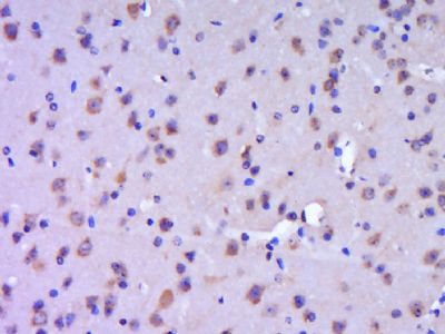
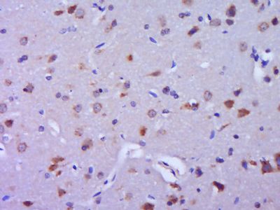
Application
| IHC-P, IHC-F, IF, ICC, E |
|---|---|
| Primary Accession | Q9C026 |
| Reactivity | Rat, Pig, Dog, Bovine |
| Host | Rabbit |
| Clonality | Polyclonal |
| Calculated MW | 79 KDa |
| Physical State | Liquid |
| Immunogen | KLH conjugated synthetic peptide derived from human TRIM9 |
| Epitope Specificity | 181-280/710 |
| Isotype | IgG |
| Purity | affinity purified by Protein A |
| Buffer | 0.01M TBS (pH7.4) with 1% BSA, 0.02% Proclin300 and 50% Glycerol. |
| SUBCELLULAR LOCATION | Cytoplasmic |
| SIMILARITY | Belongs to the TRIM/RBCC family. Contains 2 B box-type zinc fingers. Contains 1 B30.2/SPRY domain. Contains 1 COS domain. Contains 1 fibronectin type-III domain. Contains 1 RING-type zinc finger. |
| SUBUNIT | Interacts with SNAP25. |
| Post-translational modifications | Brain. Highly expressed in the cerebral cortex (at protein level). Severely decreased in the affected brain areas in Parkinson disease and dementia with Lewy bodies. |
| Important Note | This product as supplied is intended for research use only, not for use in human, therapeutic or diagnostic applications. |
| Background Descriptions | The protein encoded by this gene is a member of the tripartite motif (TRIM) family. The TRIM motif includes three zinc-binding domains, a RING, a B-box type 1 and a B-box type 2, and a coiled-coil region. The protein localizes to cytoplasmic bodies. Its function has not been identified. Alternate splicing of this gene generates two transcript variants encoding different isoforms. [provided by RefSeq, Jul 2008] |
| Gene ID | 114088 |
|---|---|
| Other Names | E3 ubiquitin-protein ligase TRIM9, 2.3.2.27, RING finger protein 91, RING-type E3 ubiquitin transferase TRIM9, Tripartite motif-containing protein 9, TRIM9, KIAA0282, RNF91 |
| Dilution | IHC-P=1:100-500,IHC-F=1:100-500,ICC=1:100-500,IF=1:100-500,ELISA=1:5000-10000 |
| Storage | Store at -20 ℃ for one year. Avoid repeated freeze/thaw cycles. When reconstituted in sterile pH 7.4 0.01M PBS or diluent of antibody the antibody is stable for at least two weeks at 2-4 ℃. |
| Name | TRIM9 |
|---|---|
| Synonyms | KIAA0282, RNF91 |
| Function | E3 ubiquitin-protein ligase which ubiquitinates itself in cooperation with an E2 enzyme UBE2D2/UBC4 and serves as a targeting signal for proteasomal degradation. May play a role in regulation of neuronal functions and may also participate in the formation or breakdown of abnormal inclusions in neurodegenerative disorders. May act as a regulator of synaptic vesicle exocytosis by controlling the availability of SNAP25 for the SNARE complex formation. |
| Cellular Location | Cytoplasm. Cell projection, dendrite. Cytoplasmic vesicle, secretory vesicle, synaptic vesicle {ECO:0000250|UniProtKB:Q91ZY8}. Synapse {ECO:0000250|UniProtKB:Q91ZY8} Cytoplasm, cytoskeleton {ECO:0000250|UniProtKB:Q91ZY8}. Note=Enriched at synaptic terminals where it exists in a soluble form and a synaptic vesicle-associated form. Associated with the cytoskeleton (By similarity). Found in proximal dendrites of pyramidal neurons in the cerebral cortex and hippocampus, and Purkinje cells in the cerebellum (PubMed:20085810). {ECO:0000250|UniProtKB:Q91ZY8, ECO:0000269|PubMed:20085810} |
| Tissue Location | Brain. Highly expressed in the cerebral cortex (at protein level). Severely decreased in the affected brain areas in Parkinson disease and dementia with Lewy bodies |
Citations (0)

Thousands of laboratories across the world have published research that depended on the performance of antibodies from Abcepta to advance their research. Check out links to articles that cite our products in major peer-reviewed journals, organized by research category.
Submit your citation using an Abcepta antibody to
info@abcepta.com, and receive a free "I Love Antibodies" mug.
info@abcepta.com, and receive a free "I Love Antibodies" mug.

Application Protocols
Provided below are standard protocols that you may find useful for product applications.
Abcepta welcomes feedback from its customers.
If you have used an Abcepta product and would like to share how it has performed, please click on the "Submit Review" button and provide the requested information. Our staff will examine and post your review and contact you if needed.
If you have any additional inquiries please email technical services at tech@abcepta.com.
$ 385.00
Cat# AP56574
Ordering Information
United States
AlbaniaAustraliaAustriaBelgiumBosnia & HerzegovinaBrazilBulgariaCanadaCentral AmericaChinaCroatiaCyprusCzech RepublicDenmarkEstoniaFinlandFranceGermanyGreeceHong KongHungaryIcelandIndiaIndonesiaIrelandIsraelItalyJapanLatviaLithuaniaLuxembourgMacedoniaMalaysiaMaltaMexicoNetherlandsNew ZealandNorwayPakistanPolandPortugalRomaniaSerbiaSingaporeSlovakiaSloveniaSouth AfricaSouth KoreaSpainSwedenSwitzerlandTaiwanTurkeyUnited KingdomUnited StatesVietnamWorldwideOthers
USA Headquarters
(888) 735-7227 / (858) 622-0099 or (858) 875-1900
Other Products
Shipping Information
Domestic orders (in stock items)
Shipped out the same day. Orders placed after 1 PM (PST) will ship out the next business day.
International orders
Contact your local distributors
