


 Foundational characteristics of cancer include proliferation, angiogenesis, migration, evasion of apoptosis, and cellular immortality. Find key markers for these cellular processes and antibodies to detect them.
 The SUMOplot™ Analysis Program predicts and scores sumoylation sites in your protein. SUMOylation is a post-translational modification involved in various cellular processes, such as nuclear-cytosolic transport, transcriptional regulation, apoptosis, protein stability, response to stress, and progression through the cell cycle.
 The Autophagy Receptor Motif Plotter predicts and scores autophagy receptor binding sites in your protein. Identifying proteins connected to this pathway is critical to understanding the role of autophagy in physiological as well as pathological processes such as development, differentiation, neurodegenerative diseases, stress, infection, and cancer.
SAFB Polyclonal Antibody
Purified Rabbit Polyclonal Antibody (Pab)
- SPECIFICATION
- CITATIONS
- PROTOCOLS
- BACKGROUND

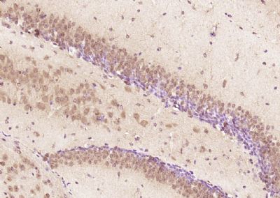
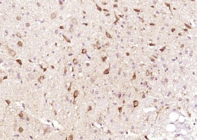
Application
| IHC-P, IHC-F, IF, ICC, E |
|---|---|
| Primary Accession | Q14151 |
| Reactivity | Rat, Pig, Bovine |
| Host | Rabbit |
| Clonality | Polyclonal |
| Calculated MW | 107 KDa |
| Physical State | Liquid |
| Immunogen | KLH conjugated synthetic peptide derived from human SAFB |
| Epitope Specificity | 551-650/953 |
| Isotype | IgG |
| Purity | affinity purified by Protein A |
| Buffer | 0.01M TBS (pH7.4) with 1% BSA, 0.02% Proclin300 and 50% Glycerol. |
| SUBCELLULAR LOCATION | Cytoplasm. Nucleus. |
| SIMILARITY | Contains 1 RRM (RNA recognition motif) domain. Contains 1 SAP domain. |
| Post-translational modifications | Phosphorylated upon DNA damage, probably by ATM or ATR. |
| Important Note | This product as supplied is intended for research use only, not for use in human, therapeutic or diagnostic applications. |
| Background Descriptions | Heterogeneous nuclear ribonucleoproteins (hnRNPs) constitute a set of polypeptides that contribute to pre-mRNA processing and transport. hnRNPs also bind heterogeneous nuclear RNA (hnRNA), the transcripts produced by RNA polymerase II. SAF-B (scaffold attachment factor B) is a nuclear matrix-associated protein that binds to matrix- or scaffold-associating regions (MARs or SARs) on DNA and interacts with RNA polymerase II and serine-/arginine-rich RNA processing factors (SR proteins). SAF-B, also designated HAP (hnRNP A1 associated protein) and HET (HSP 27-ERE-TATA-binding protein) is a proven hnRNP protein that has a speckled distribution in the nucleus and, in response to stress agents such as heat shock, is recruited to a few, large nuclear granules, called perichromatin granules. SAF-B also binds to the estrogen receptor (ER) and is expressed in several breast cancer cell lines at varying levels. Subsequently, SAF-B may play a role in breast cancer by mediating cellular proliferation and division. |
| Gene ID | 9667 |
|---|---|
| Other Names | Scaffold attachment factor B2, SAF-B2, SAFB2, KIAA0138 |
| Target/Specificity | Expressed at high levels in the CNS and at low levels in the liver. Expressed in a wide number of breast cancer cell lines. |
| Dilution | IHC-P=1:100-500,IHC-F=1:100-500,ICC=1:100-500,IF=1:100-500,ELISA=1:5000-10000 |
| Storage | Store at -20 ℃ for one year. Avoid repeated freeze/thaw cycles. When reconstituted in sterile pH 7.4 0.01M PBS or diluent of antibody the antibody is stable for at least two weeks at 2-4 ℃. |
| Name | SAFB2 |
|---|---|
| Synonyms | KIAA0138 |
| Function | Binds to scaffold/matrix attachment region (S/MAR) DNA. Can function as an estrogen receptor corepressor and can also inhibit cell proliferation. |
| Cellular Location | Cytoplasm. Nucleus. |
| Tissue Location | Expressed at high levels in the CNS and at low levels in the liver. Expressed in a wide number of breast cancer cell lines |
Research Areas
Citations (0)

Thousands of laboratories across the world have published research that depended on the performance of antibodies from Abcepta to advance their research. Check out links to articles that cite our products in major peer-reviewed journals, organized by research category.
Submit your citation using an Abcepta antibody to
info@abcepta.com, and receive a free "I Love Antibodies" mug.
info@abcepta.com, and receive a free "I Love Antibodies" mug.

Application Protocols
Provided below are standard protocols that you may find useful for product applications.
Abcepta welcomes feedback from its customers.
If you have used an Abcepta product and would like to share how it has performed, please click on the "Submit Review" button and provide the requested information. Our staff will examine and post your review and contact you if needed.
If you have any additional inquiries please email technical services at tech@abcepta.com.
$ 385.00
Cat# AP56629
Ordering Information
United States
AlbaniaAustraliaAustriaBelgiumBosnia & HerzegovinaBrazilBulgariaCanadaCentral AmericaChinaCroatiaCyprusCzech RepublicDenmarkEstoniaFinlandFranceGermanyGreeceHong KongHungaryIcelandIndiaIndonesiaIrelandIsraelItalyJapanLatviaLithuaniaLuxembourgMacedoniaMalaysiaMaltaMexicoNetherlandsNew ZealandNorwayPakistanPolandPortugalRomaniaSerbiaSingaporeSlovakiaSloveniaSouth AfricaSouth KoreaSpainSwedenSwitzerlandTaiwanTurkeyUnited KingdomUnited StatesVietnamWorldwideOthers
USA Headquarters
(888) 735-7227 / (858) 622-0099 or (858) 875-1900
Other Products
Shipping Information
Domestic orders (in stock items)
Shipped out the same day. Orders placed after 1 PM (PST) will ship out the next business day.
International orders
Contact your local distributors
