


 Foundational characteristics of cancer include proliferation, angiogenesis, migration, evasion of apoptosis, and cellular immortality. Find key markers for these cellular processes and antibodies to detect them.
 The SUMOplot™ Analysis Program predicts and scores sumoylation sites in your protein. SUMOylation is a post-translational modification involved in various cellular processes, such as nuclear-cytosolic transport, transcriptional regulation, apoptosis, protein stability, response to stress, and progression through the cell cycle.
 The Autophagy Receptor Motif Plotter predicts and scores autophagy receptor binding sites in your protein. Identifying proteins connected to this pathway is critical to understanding the role of autophagy in physiological as well as pathological processes such as development, differentiation, neurodegenerative diseases, stress, infection, and cancer.
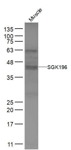
SGK196 Polyclonal Antibody
Purified Rabbit Polyclonal Antibody (Pab)
- SPECIFICATION
- CITATIONS
- PROTOCOLS
- BACKGROUND

Application
| WB, IHC-P, IHC-F, IF, ICC, E |
|---|---|
| Primary Accession | Q9H5K3 |
| Reactivity | Rat |
| Host | Rabbit |
| Clonality | Polyclonal |
| Calculated MW | 40 KDa |
| Physical State | Liquid |
| Immunogen | KLH conjugated synthetic peptide derived from human SGK196 |
| Epitope Specificity | 151-250/350 |
| Isotype | IgG |
| Purity | affinity purified by Protein A |
| Buffer | 0.01M TBS (pH7.4) with 1% BSA, 0.02% Proclin300 and 50% Glycerol. |
| SUBCELLULAR LOCATION | Cell Membrane |
| Important Note | This product as supplied is intended for research use only, not for use in human, therapeutic or diagnostic applications. |
| Background Descriptions | This gene encodes a protein that may be involved in the presentation of the laminin-binding O-linked carbohydrate chain of alpha-dystroglycan (a-DG), which forms transmembrane linkages between the extracellular matrix and the exoskeleton. Some pathogens use this O-linked carbohydrate unit for host entry. Loss of function compound heterozygous mutations in this gene were found in a human patient affected by the Walker-Warburg syndrome (WWS) phenotype. Mice lacking this gene contain misplaced neurons (heterotopia) in some regions of the brain, possibly from defects in neuronal migration. Alternative splicing of this gene results in multiple transcript variants. [provided by RefSeq, May 2013] |
| Gene ID | 84197 |
|---|---|
| Other Names | Protein O-mannose kinase, POMK, 2.7.1.183, Protein kinase-like protein SgK196, Sugen kinase 196, POMK, SGK196 |
| Dilution | WB=1:500-2000,IHC-P=1:100-500,IHC-F=1:100-500,ICC=1:100-500,IF=1:100-500,ELISA=1:5000-10000 |
| Format | 0.01M TBS(pH7.4), 0.09% (W/V) sodium azide and 50% Glyce |
| Storage | Store at -20 ℃ for one year. Avoid repeated freeze/thaw cycles. When reconstituted in sterile pH 7.4 0.01M PBS or diluent of antibody the antibody is stable for at least two weeks at 2-4 ℃. |
| Name | POMK |
|---|---|
| Synonyms | SGK196 |
| Function | Protein O-mannose kinase that specifically mediates phosphorylation at the 6-position of an O-mannose of the trisaccharide (N-acetylgalactosamine (GalNAc)-beta-1,3-N-acetylglucosamine (GlcNAc)- beta-1,4-mannose) to generate phosphorylated O-mannosyl trisaccharide (N-acetylgalactosamine-beta-1,3-N-acetylglucosamine-beta-1,4- (phosphate-6-)mannose). Phosphorylated O-mannosyl trisaccharide is a carbohydrate structure present in alpha-dystroglycan (DAG1), which is required for binding laminin G-like domain-containing extracellular proteins with high affinity. Only shows kinase activity when the GalNAc-beta-3-GlcNAc-beta-terminus is linked to the 4-position of O- mannose, suggesting that this disaccharide serves as the substrate recognition motif. |
| Cellular Location | Endoplasmic reticulum membrane; Single-pass type II membrane protein |
| Tissue Location | Highest expression is observed in brain, skeletal muscle, kidney and heart in fetal and adult tissues |
Research Areas
Citations (0)

Thousands of laboratories across the world have published research that depended on the performance of antibodies from Abcepta to advance their research. Check out links to articles that cite our products in major peer-reviewed journals, organized by research category.
Submit your citation using an Abcepta antibody to
info@abcepta.com, and receive a free "I Love Antibodies" mug.
info@abcepta.com, and receive a free "I Love Antibodies" mug.

Application Protocols
Provided below are standard protocols that you may find useful for product applications.
Abcepta welcomes feedback from its customers.
If you have used an Abcepta product and would like to share how it has performed, please click on the "Submit Review" button and provide the requested information. Our staff will examine and post your review and contact you if needed.
If you have any additional inquiries please email technical services at tech@abcepta.com.
$ 385.00
Cat# AP56656
Ordering Information
United States
AlbaniaAustraliaAustriaBelgiumBosnia & HerzegovinaBrazilBulgariaCanadaCentral AmericaChinaCroatiaCyprusCzech RepublicDenmarkEstoniaFinlandFranceGermanyGreeceHong KongHungaryIcelandIndiaIndonesiaIrelandIsraelItalyJapanLatviaLithuaniaLuxembourgMacedoniaMalaysiaMaltaMexicoNetherlandsNew ZealandNorwayPakistanPolandPortugalRomaniaSerbiaSingaporeSlovakiaSloveniaSouth AfricaSouth KoreaSpainSwedenSwitzerlandTaiwanTurkeyUnited KingdomUnited StatesVietnamWorldwideOthers
USA Headquarters
(888) 735-7227 / (858) 622-0099 or (858) 875-1900
Shipping Information
Domestic orders (in stock items)
Shipped out the same day. Orders placed after 1 PM (PST) will ship out the next business day.
International orders
Contact your local distributors
