


 Foundational characteristics of cancer include proliferation, angiogenesis, migration, evasion of apoptosis, and cellular immortality. Find key markers for these cellular processes and antibodies to detect them.
 The SUMOplot™ Analysis Program predicts and scores sumoylation sites in your protein. SUMOylation is a post-translational modification involved in various cellular processes, such as nuclear-cytosolic transport, transcriptional regulation, apoptosis, protein stability, response to stress, and progression through the cell cycle.
 The Autophagy Receptor Motif Plotter predicts and scores autophagy receptor binding sites in your protein. Identifying proteins connected to this pathway is critical to understanding the role of autophagy in physiological as well as pathological processes such as development, differentiation, neurodegenerative diseases, stress, infection, and cancer.
SPTBN3 Polyclonal Antibody
Purified Rabbit Polyclonal Antibody (Pab)
- SPECIFICATION
- CITATIONS
- PROTOCOLS
- BACKGROUND

Application
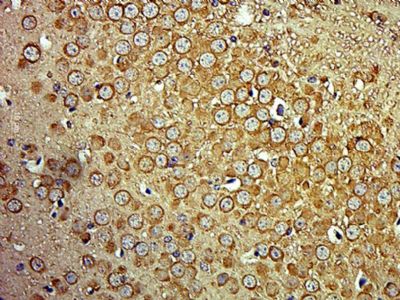
| IHC-P, IHC-F, IF, ICC, E |
|---|---|
| Primary Accession | Q9H254 |
| Reactivity | Rat, Bovine |
| Host | Rabbit |
| Clonality | Polyclonal |
| Calculated MW | 289 KDa |
| Physical State | Liquid |
| Immunogen | KLH conjugated synthetic peptide derived from human SPTBN3 |
| Epitope Specificity | 2401-2500/2564 |
| Isotype | IgG |
| Purity | affinity purified by Protein A |
| Buffer | 0.01M TBS (pH7.4) with 1% BSA, 0.02% Proclin300 and 50% Glycerol. |
| SUBCELLULAR LOCATION | Cytoplasm > cytoskeleton. Cytoplasm > cell cortex. |
| SIMILARITY | Belongs to the spectrin family.Contains 2 CH (calponin-homology) domains. Contains 1 PH domain. Contains 18 spectrin repeats. |
| Important Note | This product as supplied is intended for research use only, not for use in human, therapeutic or diagnostic applications. |
| Background Descriptions | Spectrin, an actin binding protein that is a major component of the cytoskeletal superstructure of the erythrocyte plasma membrane, is essential in determining the properties of the membrane including its shape and deformability. Spectrins function as membrane organizers and stabilizers, composed of nonhomologous Alpha and beta chains, which aggregate side-to-side in an antiparallel fashion to form dimers, tetramers, and higher polymers. The spectrin tetramers in erythrocytes act as barriers to lateral diffusion, but spectrin dimers seem to lack this function. Spectrin beta IV is a non-erythrocytic member of the beta-spectrin family. It is expressed in brain and pancreatic islets and localizes to the nuclear matrix, cytoplasmic vesicles and PML nuclear bodies. Spectrin beta IV is a 2,564 amino acid protein with four isoforms due to alternative splicing events. |
| Gene ID | 57731 |
|---|---|
| Other Names | Spectrin beta chain, non-erythrocytic 4, Beta-IV spectrin, Spectrin, non-erythroid beta chain 3, SPTBN4, KIAA1642, SPTBN3 |
| Target/Specificity | Abundantly expressed in brain and pancreatic islets. |
| Dilution | IHC-P=1:100-500,IHC-F=1:100-500,ICC=1:100-500,IF=1:100-500,ELISA=1:5000-10000 |
| Storage | Store at -20 ℃ for one year. Avoid repeated freeze/thaw cycles. When reconstituted in sterile pH 7.4 0.01M PBS or diluent of antibody the antibody is stable for at least two weeks at 2-4 ℃. |
| Name | SPTBN4 |
|---|---|
| Synonyms | KIAA1642, SPTBN3 |
| Cellular Location | Cytoplasm, cytoskeleton. Cytoplasm, cell cortex |
| Tissue Location | Expressed in skeletal muscle at the sarcolemma and in the muscle capillaries (at protein level) (PubMed:28540413) Abundantly expressed in brain and pancreatic islets (PubMed:11086001) |
Research Areas
Citations (0)

Thousands of laboratories across the world have published research that depended on the performance of antibodies from Abcepta to advance their research. Check out links to articles that cite our products in major peer-reviewed journals, organized by research category.
Submit your citation using an Abcepta antibody to
info@abcepta.com, and receive a free "I Love Antibodies" mug.
info@abcepta.com, and receive a free "I Love Antibodies" mug.

Application Protocols
Provided below are standard protocols that you may find useful for product applications.
Abcepta welcomes feedback from its customers.
If you have used an Abcepta product and would like to share how it has performed, please click on the "Submit Review" button and provide the requested information. Our staff will examine and post your review and contact you if needed.
If you have any additional inquiries please email technical services at tech@abcepta.com.
$ 385.00
Cat# AP56774
Ordering Information
United States
AlbaniaAustraliaAustriaBelgiumBosnia & HerzegovinaBrazilBulgariaCanadaCentral AmericaChinaCroatiaCyprusCzech RepublicDenmarkEstoniaFinlandFranceGermanyGreeceHong KongHungaryIcelandIndiaIndonesiaIrelandIsraelItalyJapanLatviaLithuaniaLuxembourgMacedoniaMalaysiaMaltaMexicoNetherlandsNew ZealandNorwayPakistanPolandPortugalRomaniaSerbiaSingaporeSlovakiaSloveniaSouth AfricaSouth KoreaSpainSwedenSwitzerlandTaiwanTurkeyUnited KingdomUnited StatesVietnamWorldwideOthers
USA Headquarters
(888) 735-7227 / (858) 622-0099 or (858) 875-1900
Shipping Information
Domestic orders (in stock items)
Shipped out the same day. Orders placed after 1 PM (PST) will ship out the next business day.
International orders
Contact your local distributors
