


 Foundational characteristics of cancer include proliferation, angiogenesis, migration, evasion of apoptosis, and cellular immortality. Find key markers for these cellular processes and antibodies to detect them.
 The SUMOplot™ Analysis Program predicts and scores sumoylation sites in your protein. SUMOylation is a post-translational modification involved in various cellular processes, such as nuclear-cytosolic transport, transcriptional regulation, apoptosis, protein stability, response to stress, and progression through the cell cycle.
 The Autophagy Receptor Motif Plotter predicts and scores autophagy receptor binding sites in your protein. Identifying proteins connected to this pathway is critical to understanding the role of autophagy in physiological as well as pathological processes such as development, differentiation, neurodegenerative diseases, stress, infection, and cancer.
MSRB2 Polyclonal Antibody
Purified Rabbit Polyclonal Antibody (Pab)
- SPECIFICATION
- CITATIONS
- PROTOCOLS
- BACKGROUND

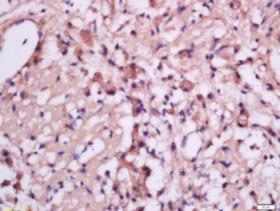
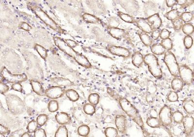
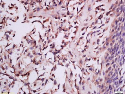
Application
| IHC-P, IHC-F, IF, ICC, E |
|---|---|
| Primary Accession | Q9Y3D2 |
| Reactivity | Rat, Dog |
| Host | Rabbit |
| Clonality | Polyclonal |
| Calculated MW | 20 KDa |
| Physical State | Liquid |
| Immunogen | KLH conjugated synthetic peptide derived from human MSRB2 |
| Epitope Specificity | 31-130/182 |
| Isotype | IgG |
| Purity | affinity purified by Protein A |
| Buffer | 0.01M TBS (pH7.4) with 1% BSA, 0.02% Proclin300 and 50% Glycerol. |
| SUBCELLULAR LOCATION | Mitochondrion. |
| SIMILARITY | Belongs to the MsrB Met sulfoxide reductase family. |
| Important Note | This product as supplied is intended for research use only, not for use in human, therapeutic or diagnostic applications. |
| Background Descriptions | Methionine is one of the most readily oxidized essential amino acids and an intermediate in the biosynthesis of cysteine, carnitine, taurine, lecithin, phosphatidylcholine and other phospholipids. In its oxidative state, Methionine is regulated in vivo by methionine sulfoxide reductases (Msr). MsrB2 is a 182 amino acid mitochondrial protein that is ubiquitously expressed. Belonging to the MsrB Met sulfoxide reductase family, MsrB2 acts as a catalyst for the reduction of free and protein-bound methionine sulfoxide to methionine. Upon oxidative stress, MsrB2 is suggested to play a role in the preservation of mitochondrial integrity by decreasing the intracellular reactive oxygen species build-up through its scavenging role, hence contributing to cell survival and protein maintenance. MsrB2 utilizes zinc ions, one per subunit, as cofactors. |
| Gene ID | 22921 |
|---|---|
| Other Names | Methionine-R-sulfoxide reductase B2, mitochondrial, MsrB2, 1.8.4.12, MSRB2, CBS-1, MSRB |
| Target/Specificity | Ubiquitous. Detected in retina, ocular ciliary body, skeletal muscle, heart, colon, bone marrow, cerebellum, small intestine, fetal brain, fetal liver, kidney, spinal cord, lung, placenta and prostate. |
| Dilution | IHC-P=1:100-500,IHC-F=1:100-500,ICC=1:100-500,IF=1:100-500,ELISA=1:5000-10000 |
| Storage | Store at -20 ℃ for one year. Avoid repeated freeze/thaw cycles. When reconstituted in sterile pH 7.4 0.01M PBS or diluent of antibody the antibody is stable for at least two weeks at 2-4 ℃. |
| Name | MSRB2 |
|---|---|
| Synonyms | CBS-1, MSRB |
| Function | Methionine-sulfoxide reductase that specifically reduces methionine (R)-sulfoxide back to methionine. While in many cases, methionine oxidation is the result of random oxidation following oxidative stress, methionine oxidation is also a post-translational modification that takes place on specific residue. Upon oxidative stress, may play a role in the preservation of mitochondrial integrity by decreasing the intracellular reactive oxygen species build-up through its scavenging role, hence contributing to cell survival and protein maintenance. |
| Cellular Location | Mitochondrion |
| Tissue Location | Ubiquitous. Detected in retina, ocular ciliary body, skeletal muscle, heart, colon, bone marrow, cerebellum, small intestine, fetal brain, fetal liver, kidney, spinal cord, lung, placenta and prostate. |
Research Areas
Citations (0)

Thousands of laboratories across the world have published research that depended on the performance of antibodies from Abcepta to advance their research. Check out links to articles that cite our products in major peer-reviewed journals, organized by research category.
Submit your citation using an Abcepta antibody to
info@abcepta.com, and receive a free "I Love Antibodies" mug.
info@abcepta.com, and receive a free "I Love Antibodies" mug.

Application Protocols
Provided below are standard protocols that you may find useful for product applications.
Abcepta welcomes feedback from its customers.
If you have used an Abcepta product and would like to share how it has performed, please click on the "Submit Review" button and provide the requested information. Our staff will examine and post your review and contact you if needed.
If you have any additional inquiries please email technical services at tech@abcepta.com.
$ 385.00
Cat# AP56872
Ordering Information
United States
AlbaniaAustraliaAustriaBelgiumBosnia & HerzegovinaBrazilBulgariaCanadaCentral AmericaChinaCroatiaCyprusCzech RepublicDenmarkEstoniaFinlandFranceGermanyGreeceHong KongHungaryIcelandIndiaIndonesiaIrelandIsraelItalyJapanLatviaLithuaniaLuxembourgMacedoniaMalaysiaMaltaMexicoNetherlandsNew ZealandNorwayPakistanPolandPortugalRomaniaSerbiaSingaporeSlovakiaSloveniaSouth AfricaSouth KoreaSpainSwedenSwitzerlandTaiwanTurkeyUnited KingdomUnited StatesVietnamWorldwideOthers
USA Headquarters
(888) 735-7227 / (858) 622-0099 or (858) 875-1900
Other Products
Shipping Information
Domestic orders (in stock items)
Shipped out the same day. Orders placed after 1 PM (PST) will ship out the next business day.
International orders
Contact your local distributors
