


 Foundational characteristics of cancer include proliferation, angiogenesis, migration, evasion of apoptosis, and cellular immortality. Find key markers for these cellular processes and antibodies to detect them.
 The SUMOplot™ Analysis Program predicts and scores sumoylation sites in your protein. SUMOylation is a post-translational modification involved in various cellular processes, such as nuclear-cytosolic transport, transcriptional regulation, apoptosis, protein stability, response to stress, and progression through the cell cycle.
 The Autophagy Receptor Motif Plotter predicts and scores autophagy receptor binding sites in your protein. Identifying proteins connected to this pathway is critical to understanding the role of autophagy in physiological as well as pathological processes such as development, differentiation, neurodegenerative diseases, stress, infection, and cancer.
MAFG Polyclonal Antibody
Purified Rabbit Polyclonal Antibody (Pab)
- SPECIFICATION
- CITATIONS
- PROTOCOLS
- BACKGROUND

Application
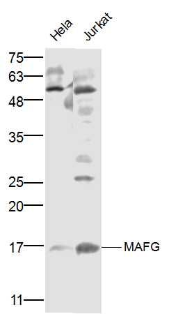
| WB, IHC-P, IHC-F, IF, ICC |
|---|---|
| Primary Accession | O15525 |
| Reactivity | Rat, Pig, Dog, Bovine |
| Host | Rabbit |
| Clonality | Polyclonal |
| Calculated MW | 18 KDa |
| Physical State | Liquid |
| Immunogen | KLH conjugated synthetic peptide derived from human MAFG |
| Epitope Specificity | 1-100/162 |
| Isotype | IgG |
| Purity | affinity purified by Protein A |
| Buffer | 0.01M TBS (pH7.4) with 1% BSA, 0.02% Proclin300 and 50% Glycerol. |
| SUBCELLULAR LOCATION | Nucleus. |
| SIMILARITY | Belongs to the bZIP family. Maf subfamily. Contains 1 bZIP domain. |
| Post-translational modifications | Acetylated in erythroid cells by CREB-binding protein (CBP). Acetylation augments the DNA-binding activity of NFE2, but has no effect on binding NFE2. Sumoylation at Lys-14 is required for active transcriptional repression. |
| Important Note | This product as supplied is intended for research use only, not for use in human, therapeutic or diagnostic applications. |
| Background Descriptions | Globin gene expression is regulated through nuclear factor erythroid-2 (NFE2) elements located in enhancer-like locus control regions positioned many kb upstream of alpha- and beta-gene clusters (summarized by Blank et al., 1997 [PubMed 9166829]). NFE2 DNA-binding activity consists of a heterodimer containing a ubiquitous small Maf protein (MafF, MIM 604877; MafG; or MafK, MIM 600197) and the tissue-restricted protein p45 NFE2 (MIM 601490). Both subunits are members of the activator protein-1-like superfamily of basic leucine zipper (bZIP) proteins (see MIM 165160).[supplied by OMIM, Mar 2010] |
| Gene ID | 4097 |
|---|---|
| Other Names | Transcription factor MafG, V-maf musculoaponeurotic fibrosarcoma oncogene homolog G, hMAF, MAFG |
| Target/Specificity | Highly expressed in skeletal muscle. Also expressed in heart and brain. |
| Dilution | WB=1:500-2000,IHC-P=1:100-500,IHC-F=1:100-500,ICC=1:100-500,IF=1:100-500 |
| Storage | Store at -20 ℃ for one year. Avoid repeated freeze/thaw cycles. When reconstituted in sterile pH 7.4 0.01M PBS or diluent of antibody the antibody is stable for at least two weeks at 2-4 ℃. |
| Name | MAFG |
|---|---|
| Function | Since they lack a putative transactivation domain, the small Mafs behave as transcriptional repressors when they dimerize among themselves (PubMed:11154691). However, they seem to serve as transcriptional activators by dimerizing with other (usually larger) basic-zipper proteins, such as NFE2, NFE2L1 and NFE2L2, and recruiting them to specific DNA-binding sites (PubMed:11154691, PubMed:8932385, PubMed:9421508). Small Maf proteins heterodimerize with Fos and may act as competitive repressors of the NFE2L2 transcription factor (PubMed:11154691). Transcription factor, component of erythroid- specific transcription factor NFE2L2 (PubMed:11154691). Activates globin gene expression when associated with NFE2L2 (PubMed:11154691). May be involved in signal transduction of extracellular H(+) (By similarity). |
| Cellular Location | Nucleus {ECO:0000255|PROSITE-ProRule:PRU00978, ECO:0000269|PubMed:11154691} |
| Tissue Location | Highly expressed in skeletal muscle. Also expressed in heart and brain |
Research Areas
Citations (0)

Thousands of laboratories across the world have published research that depended on the performance of antibodies from Abcepta to advance their research. Check out links to articles that cite our products in major peer-reviewed journals, organized by research category.
Submit your citation using an Abcepta antibody to
info@abcepta.com, and receive a free "I Love Antibodies" mug.
info@abcepta.com, and receive a free "I Love Antibodies" mug.

Application Protocols
Provided below are standard protocols that you may find useful for product applications.
Abcepta welcomes feedback from its customers.
If you have used an Abcepta product and would like to share how it has performed, please click on the "Submit Review" button and provide the requested information. Our staff will examine and post your review and contact you if needed.
If you have any additional inquiries please email technical services at tech@abcepta.com.
$ 385.00
Cat# AP57180
Ordering Information
United States
AlbaniaAustraliaAustriaBelgiumBosnia & HerzegovinaBrazilBulgariaCanadaCentral AmericaChinaCroatiaCyprusCzech RepublicDenmarkEstoniaFinlandFranceGermanyGreeceHong KongHungaryIcelandIndiaIndonesiaIrelandIsraelItalyJapanLatviaLithuaniaLuxembourgMacedoniaMalaysiaMaltaMexicoNetherlandsNew ZealandNorwayPakistanPolandPortugalRomaniaSerbiaSingaporeSlovakiaSloveniaSouth AfricaSouth KoreaSpainSwedenSwitzerlandTaiwanTurkeyUnited KingdomUnited StatesVietnamWorldwideOthers
USA Headquarters
(888) 735-7227 / (858) 622-0099 or (858) 875-1900
Shipping Information
Domestic orders (in stock items)
Shipped out the same day. Orders placed after 1 PM (PST) will ship out the next business day.
International orders
Contact your local distributors
