


 Foundational characteristics of cancer include proliferation, angiogenesis, migration, evasion of apoptosis, and cellular immortality. Find key markers for these cellular processes and antibodies to detect them.
 The SUMOplot™ Analysis Program predicts and scores sumoylation sites in your protein. SUMOylation is a post-translational modification involved in various cellular processes, such as nuclear-cytosolic transport, transcriptional regulation, apoptosis, protein stability, response to stress, and progression through the cell cycle.
 The Autophagy Receptor Motif Plotter predicts and scores autophagy receptor binding sites in your protein. Identifying proteins connected to this pathway is critical to understanding the role of autophagy in physiological as well as pathological processes such as development, differentiation, neurodegenerative diseases, stress, infection, and cancer.
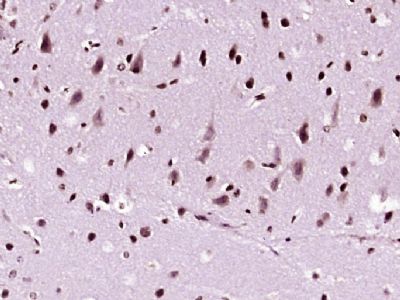
NDST4 Polyclonal Antibody
Purified Rabbit Polyclonal Antibody (Pab)
- SPECIFICATION
- CITATIONS
- PROTOCOLS
- BACKGROUND

Application
| IHC-P, IHC-F, IF, ICC, E |
|---|---|
| Primary Accession | Q9H3R1 |
| Reactivity | Rat, Pig, Dog, Bovine |
| Host | Rabbit |
| Clonality | Polyclonal |
| Calculated MW | 101 KDa |
| Physical State | Liquid |
| Immunogen | KLH conjugated synthetic peptide derived from human NDST4 |
| Epitope Specificity | 101-200/872 |
| Isotype | IgG |
| Purity | affinity purified by Protein A |
| Buffer | 0.01M TBS (pH7.4) with 1% BSA, 0.02% Proclin300 and 50% Glycerol. |
| SUBCELLULAR LOCATION | Golgi apparatus membrane; Single-pass type II membrane protein |
| SIMILARITY | Belongs to the sulfotransferase 1 family. NDST subfamily. |
| SUBUNIT | Monomer |
| Important Note | This product as supplied is intended for research use only, not for use in human, therapeutic or diagnostic applications. |
| Background Descriptions | The N-deacetylation and N-sulfation of N-acetylglucosamine residues in heparan sulfate and heparin initiate a set of biochemical reactions, which lead to the synthesis of oligosaccharide sequences that have specific ligand binding properties (1). These reactions are catalyzed by the monomeric enzymes GlcNAc Ndeacetylase/N-sulfotransferases (NDSTs), which have two catalytic activities (1). Multiple NDST isozymes have been identified, each having unique tissue distribution and enzymatic properties (2). Phylogenetic data suggests that NDST1-4 evolved from a common ancestral gene, which diverged to give rise to two subtypes, NDST1/2 and NDST3/4 (2). NDST1, which maps to human chromosome 5q32-q33.1, shares the most homology with NDST2, which maps to human chromosome 10q22 (3,4). The least conserved amino acids between these two enzymes are found in the N-terminus/putative transmembrane regions (3). The human NDST3 and NDST4 genes are closely linked on chromosome 4, mapping to chromosome 4q25-26 and 4q26-27, respectively (2,5,6). RT-PCR analysis of various mouse tissues reveals a restricted pattern of NDST3 and NDST4 mRNA expression when compared with that of NDST1 and NDST2, which are abundantly and ubiquitously expressed (2). |
| Gene ID | 64579 |
|---|---|
| Other Names | Bifunctional heparan sulfate N-deacetylase/N-sulfotransferase 4, 2.8.2.8, Glucosaminyl N-deacetylase/N-sulfotransferase 4, NDST-4, N-heparan sulfate sulfotransferase 4, N-HSST 4, Heparan sulfate N-deacetylase 4, 3.-.-.-, Heparan sulfate N-sulfotransferase 4, 2.8.2.-, NDST4, HSST4 |
| Dilution | IHC-P=1:100-500,IHC-F=1:100-500,ICC=1:100-500,IF=1:100-500,ELISA=1:5000-10000 |
| Storage | Store at -20 ℃ for one year. Avoid repeated freeze/thaw cycles. When reconstituted in sterile pH 7.4 0.01M PBS or diluent of antibody the antibody is stable for at least two weeks at 2-4 ℃. |
| Name | NDST4 |
|---|---|
| Synonyms | HSST4 |
| Function | Essential bifunctional enzyme that catalyzes both the N- deacetylation and the N-sulfation of glucosamine (GlcNAc) of the glycosaminoglycan in heparan sulfate. Modifies the GlcNAc-GlcA disaccharide repeating sugar backbone to make N-sulfated heparosan, a prerequisite substrate for later modifications in heparin biosynthesis. Has low deacetylase activity but high sulfotransferase activity (By similarity). |
| Cellular Location | Golgi apparatus membrane; Single- pass type II membrane protein |
Research Areas
Citations (0)

Thousands of laboratories across the world have published research that depended on the performance of antibodies from Abcepta to advance their research. Check out links to articles that cite our products in major peer-reviewed journals, organized by research category.
Submit your citation using an Abcepta antibody to
info@abcepta.com, and receive a free "I Love Antibodies" mug.
info@abcepta.com, and receive a free "I Love Antibodies" mug.

Application Protocols
Provided below are standard protocols that you may find useful for product applications.
Abcepta welcomes feedback from its customers.
If you have used an Abcepta product and would like to share how it has performed, please click on the "Submit Review" button and provide the requested information. Our staff will examine and post your review and contact you if needed.
If you have any additional inquiries please email technical services at tech@abcepta.com.
$ 385.00
Cat# AP57380
Ordering Information
United States
AlbaniaAustraliaAustriaBelgiumBosnia & HerzegovinaBrazilBulgariaCanadaCentral AmericaChinaCroatiaCyprusCzech RepublicDenmarkEstoniaFinlandFranceGermanyGreeceHong KongHungaryIcelandIndiaIndonesiaIrelandIsraelItalyJapanLatviaLithuaniaLuxembourgMacedoniaMalaysiaMaltaMexicoNetherlandsNew ZealandNorwayPakistanPolandPortugalRomaniaSerbiaSingaporeSlovakiaSloveniaSouth AfricaSouth KoreaSpainSwedenSwitzerlandTaiwanTurkeyUnited KingdomUnited StatesVietnamWorldwideOthers
USA Headquarters
(888) 735-7227 / (858) 622-0099 or (858) 875-1900
Shipping Information
Domestic orders (in stock items)
Shipped out the same day. Orders placed after 1 PM (PST) will ship out the next business day.
International orders
Contact your local distributors
