


 Foundational characteristics of cancer include proliferation, angiogenesis, migration, evasion of apoptosis, and cellular immortality. Find key markers for these cellular processes and antibodies to detect them.
 The SUMOplot™ Analysis Program predicts and scores sumoylation sites in your protein. SUMOylation is a post-translational modification involved in various cellular processes, such as nuclear-cytosolic transport, transcriptional regulation, apoptosis, protein stability, response to stress, and progression through the cell cycle.
 The Autophagy Receptor Motif Plotter predicts and scores autophagy receptor binding sites in your protein. Identifying proteins connected to this pathway is critical to understanding the role of autophagy in physiological as well as pathological processes such as development, differentiation, neurodegenerative diseases, stress, infection, and cancer.
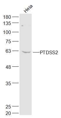
PTDSS2 Polyclonal Antibody
Purified Rabbit Polyclonal Antibody (Pab)
- SPECIFICATION
- CITATIONS
- PROTOCOLS
- BACKGROUND

Application
| WB, IHC-P, IHC-F, IF, ICC, E |
|---|---|
| Primary Accession | Q9BVG9 |
| Reactivity | Rat, Dog |
| Host | Rabbit |
| Clonality | Polyclonal |
| Calculated MW | 56 KDa |
| Physical State | Liquid |
| Immunogen | KLH conjugated synthetic peptide derived from human PTDSS2 |
| Epitope Specificity | 1-100/487 |
| Isotype | IgG |
| Purity | affinity purified by Protein A |
| Buffer | 0.01M TBS (pH7.4) with 1% BSA, 0.02% Proclin300 and 50% Glycerol. |
| SUBCELLULAR LOCATION | Membrane. |
| SIMILARITY | Belongs to the phosphatidyl serine synthase family. |
| Important Note | This product as supplied is intended for research use only, not for use in human, therapeutic or diagnostic applications. |
| Background Descriptions | Phosphatidylserine (PS) accounts for 5 to 10% of cell membrane phospholipids. In addition to its role as a structural component, PS is involved in cell signaling, blood coagulation, and apoptosis. PS is synthesized by a calcium-dependent base-exchange reaction catalyzed by PS synthases (EC 2.7.8.8), like PTDSS2, that exchange L-serine for the polar head group of phosphatidylcholine (PC) or phosphatidylethanolamine (PE) (Sturbois-Balcerzak et al., 2001 [PubMed 11084049]).[supplied by OMIM, May 2009] |
| Gene ID | 81490 |
|---|---|
| Other Names | Phosphatidylserine synthase 2, PSS-2, PtdSer synthase 2, 2.7.8.29, Serine-exchange enzyme II, PTDSS2, PSS2 |
| Dilution | WB=1:500-2000,IHC-P=1:100-500,IHC-F=1:100-500,ICC=1:100-500,IF=1:100-500,ELISA=1:5000-10000 |
| Storage | Store at -20 ℃ for one year. Avoid repeated freeze/thaw cycles. When reconstituted in sterile pH 7.4 0.01M PBS or diluent of antibody the antibody is stable for at least two weeks at 2-4 ℃. |
| Name | PTDSS2 |
|---|---|
| Synonyms | PSS2 |
| Function | Catalyzes a base-exchange reaction in which the polar head group of phosphatidylethanolamine (PE) or phosphatidylcholine (PC) is replaced by L-serine (PubMed:19014349). Catalyzes the conversion of phosphatatidylethanolamine and does not act on phosphatidylcholine (PubMed:19014349). Can utilize both phosphatidylethanolamine (PE) plasmalogen and diacyl PE as substrate and the latter is six times better utilized, indicating the importance of an ester linkage at the sn-1 position (By similarity). Although it shows no sn-1 fatty acyl preference, exhibits significant preference towards docosahexaenoic acid (22:6n-3) compared with 18:1 or 20:4 at the sn-2 position (By similarity). |
| Cellular Location | Endoplasmic reticulum membrane {ECO:0000250|UniProtKB:Q9Z1X2}; Multi-pass membrane protein. Note=Highly enriched in the mitochondria-associated membrane (MAM). {ECO:0000250|UniProtKB:Q9Z1X2} |
Research Areas
Citations (0)

Thousands of laboratories across the world have published research that depended on the performance of antibodies from Abcepta to advance their research. Check out links to articles that cite our products in major peer-reviewed journals, organized by research category.
Submit your citation using an Abcepta antibody to
info@abcepta.com, and receive a free "I Love Antibodies" mug.
info@abcepta.com, and receive a free "I Love Antibodies" mug.

Application Protocols
Provided below are standard protocols that you may find useful for product applications.
Abcepta welcomes feedback from its customers.
If you have used an Abcepta product and would like to share how it has performed, please click on the "Submit Review" button and provide the requested information. Our staff will examine and post your review and contact you if needed.
If you have any additional inquiries please email technical services at tech@abcepta.com.
$ 385.00
Cat# AP57587
Ordering Information
United States
AlbaniaAustraliaAustriaBelgiumBosnia & HerzegovinaBrazilBulgariaCanadaCentral AmericaChinaCroatiaCyprusCzech RepublicDenmarkEstoniaFinlandFranceGermanyGreeceHong KongHungaryIcelandIndiaIndonesiaIrelandIsraelItalyJapanLatviaLithuaniaLuxembourgMacedoniaMalaysiaMaltaMexicoNetherlandsNew ZealandNorwayPakistanPolandPortugalRomaniaSerbiaSingaporeSlovakiaSloveniaSouth AfricaSouth KoreaSpainSwedenSwitzerlandTaiwanTurkeyUnited KingdomUnited StatesVietnamWorldwideOthers
USA Headquarters
(888) 735-7227 / (858) 622-0099 or (858) 875-1900
Other Products
Shipping Information
Domestic orders (in stock items)
Shipped out the same day. Orders placed after 1 PM (PST) will ship out the next business day.
International orders
Contact your local distributors
